- Head and Neck
- Dental Anatomy
- Upper Extremity
- Thorax
- Abdomen
- Spine and Back
- Pelvis
- Lower Extremity
- Organ Systems
- Anatomical terminology
- Skeletal system
- Skull
- Neurocranium
- Frontal bone
- Sphenoid bone
- Temporal bone
- Ethmoid bone
- Occipital bone
- Parietal bone
- Viscerocranium
- Lacrimal bone
- Nasal bone
- Zygomatic bone
- Palatine bone
- Maxilla
- Inferior nasal concha
- Vomer
- Mandible
- Hyoid bone
- Auditory ossicles
- Sutures of skull
- Topography of skull
- Cranial base
- Internal cranial base
- Anterior cranial fossa
- Middle cranial fossa
- Posterior cranial fossa
- External cranial base
- Temporal fossa
- Infratemporal fossa
- Pterygopalatine fossa
- Orbit
- Nasal cavity
- Paranasal sinuses
- Maxillary sinus
- Frontal sinus
- Sphenoidal sinus
- Ethmoidal air cells
- Skeleton of trunk
- Rib cage
- Ribs
- Sternum
- Thoracic vertebrae
- Spine
- Cervical vertebrae
- Thoracic vertebrae
- Lumbar vertebrae
- Sacrum
- Coccyx
- Skeleton of upper limb
- Bones of shoulder girdle
- Clavicle
- Scapula
- Humerus
- Bones of forearm
- Radius
- Ulna
- Bones of hand
- Carpal bones
- Metacarpal bones
- Phalanges of hand
- Skeleton of lower limb
- Pelvis
- Hip bone
- Pubic bone
- Ischium
- Ilium
- Sacrum
- Coccyx
- Femur
- Patella
- Bones of leg
- Tibia
- Fibula
- Bones of foot
- Tarsal bones
- Talus
- Calcaneus
- Cuboid bone
- Navicular bone
- Cuneiform bones
- Metatarsal bones
- Phalanges of foot
- Joints
- Classification of joints
- Joints of skull
- Temporomandibular joint
- Sutures of skull
- Joints of spine
- Anterior longitudinal ligament
- Posterior longitudinal ligament
- Supraspinous and nuchal ligaments
- Ligamenta flava
- Intervertebral discs
- Atlanto-occipital joint
- Atlanto-axial joint
- Facet (zygapophyseal) joints
- Lumbosacral joint
- Sacrococcygeal joint
- Joints of lower limb
- Joints of pelvis
- Sacrotuberous ligament
- Sacrospinous ligament
- Obturator membrane
- Pubic symphysis
- Sacroiliac joint
- Hip joint
- Knee joint
- Tibiofibular joints
- Superior tibiofibular joint
- Inferior tibiofibular joint
- Joints of foot
- Ankle joint
- Intertarsal joints
- Talocalcaneonavicular joint
- Calcaneocuboid joint
- Transverse tarsal joint (Chopart's joint)
- Cuneonavicular joint
- Subtalar joint
- Tarsometatarsal joints (Lisfranc's joint)
- Intermetatarsal joints
- Metatarsophalangeal joints
- Interphalangeal joints of foot
- Muscles
- Head muscles
- Extraocular muscles
- Superior rectus
- Inferior rectus
- Medial rectus
- Lateral rectus
- Superior oblique
- Inferior oblique
- Levator palpebrae superioris
- Facial muscles
- Occipitofrontalis
- Occipital belly of occipitofrontalis
- Frontal belly of occipitofrontalis
- Corrugator supercilii
- Depressor supercilii
- Orbicularis oculi
- Orbital part of orbicularis oculi
- Palpebral part of orbicularis oculi
- Lacrimal part of orbicularis oculi
- Buccinator
- Orbicularis oris
- Mentalis
- Depressor anguli oris
- Depressor labii inferioris
- Levator anguli oris
- Levator labii superioris
- Risorius
- Zygomaticus major
- Zygomaticus minor
- Levator labii superioris alaeque nasi
- Nasalis
- Alar nasalis
- Transverse nasalis
- Procerus
- Depressor septi nasi
- Compressor narium minor
- Dilator naris anterior
- Muscles of mastication
- Temporalis
- Masseter
- Lateral pterygoid
- Medial pterygoid
- Neck muscles
- Superficial neck muscles
- Sternocleidomastoid
- Platysma
- Scalene muscles
- Anterior scalene
- Middle scalene
- Posterior scalene
- Suprahyoid muscles
- Mylohyoid
- Digastric
- Anterior belly of digastric
- Posterior belly of digastric
- Stylohyoid
- Geniohyoid
- Infrahyoid muscles
- Sternohyoid
- Sternothyroid
- Thyrohyoid
- Omohyoid
- Prevertebral muscles
- Longus capitis
- Longus colli
- Rectus capitis anterior
- Rectus capitis lateralis
- Suboccipital muscles
- Rectus capitis posterior minor
- Rectus capitis posterior major
- Obliquus capitis superior
- Obliquus capitis inferior
- Muscles of upper limb
- Muscles of pectoral girdle
- Pectoralis major
- Muscles of shoulder region
- Deltoid
- Teres major
- Rotator cuff
- Supraspinatus
- Infraspinatus
- Teres minor
- Subscapularis
- Muscles of upper arm
- Anterior compartment
- Coracobrachialis
- Brachialis
- Biceps brachii
- Posterior compartment
- Triceps brachii
- Anconeus
- Muscles of forearm
- Anterior compartment
- Pronator teres
- Flexor carpi radialis
- Palmaris longus
- Flexor carpi ulnaris
- Flexor digitorum superficialis
- Flexor digitorum profundus
- Flexor pollicis longus
- Pronator quadratus
- Lateral compartment
- Brachioradialis
- Extensor carpi radialis longus
- Extensor carpi radialis brevis
- Posterior compartment
- Extensor digitorum
- Extensor digiti minimi
- Extensor carpi ulnaris
- Abductor pollicis longus
- Extensor pollicis brevis
- Extensor pollicis longus
- Extensor indicis
- Supinator
- Muscles of hand
- Medial group (Muscles of little finger)
- Abductor digiti minimi of hand
- Flexor digiti minimi brevis of hand
- Opponens digiti minimi of hand
- Palmaris brevis
- Middle group of hand muscles
- Lumbricals of hand
- Palmar interossei
- Dorsal interossei of hand
- Lateral group (Muscles of thumb)
- Abductor pollicis brevis
- Flexor pollicis brevis
- Opponens pollicis
- Adductor pollicis
- Thoracic muscles
- Diaphragm
- Muscles of back
- Superficial back muscles
- Trapezius
- Latissimus dorsi
- Rhomboid muscles
- Levator scapulae
- Intermediate back muscles
- Serratus posterior superior
- Serratus posterior inferior
- Deep back muscles
- Superficial layer
- Splenius capitis
- Splenius cervicis
- Intermediate layer (Erector Spinae)
- Spinalis
- Longissimus
- Iliocostalis
- Deep layer (Transversospinales)
- Rotatores
- Multifidus
- Semispinalis
- Deepest layer
- Interspinales
- Intertransversarii
- Levatores costarum
- Muscles of lower limb
- Pelvic muscles
- Iliopsoas
- Iliacus
- Psoas major
- Psoas minor
- Gluteus maximus
- Gluteus medius
- Gluteus minimus
- Tensor fasciae latae
- Piriformis
- Obturator internus
- Obturator externus
- Superior gemellus
- Inferior gemellus
- Quadratus femoris
- Muscles of thigh
- Anterior compartment
- Quadriceps femoris
- Rectus femoris
- Vastus medialis
- Vastus intermedius
- Vastus lateralis
- Sartorius
- Medial compartment
- Pectineus
- Adductor brevis
- Adductor longus
- Adductor magnus
- Gracilis
- Posterior compartment
- Semimembranosus
- Semitendinosus
- Biceps femoris
- Muscles of leg
- Anterior compartment
- Tibialis anterior
- Extensor hallucis longus
- Extensor digitorum longus
- Lateral compartment
- Peroneus longus
- Peroneus brevis
- Posterior compartment
- Superficial layer
- Triceps surae
- Gastrocnemius
- Soleus
- Plantaris
- Deep layer
- Popliteus
- Tibialis posterior
- Flexor digitorum longus
- Flexor hallucis longus
- Muscles of foot
- Dorsal muscles of foot
- Extensor digitorum brevis
- Extensor hallucis brevis
- Plantar muscles of foot
- Medial group (Muscles of big toe)
- Abductor hallucis
- Flexor hallucis brevis
- Adductor hallucis
- Middle group of foot muscles
- Flexor digitorum brevis
- Quadratus plantae
- Lumbricals of foot
- Plantar interossei
- Dorsal interossei of foot
- Lateral group (Muscles of little toe)
- Abductor digiti minimi of foot
- Flexor digiti minimi brevis of foot
- Opponens digiti minimi of foot
- Heart
- Blood vessels
- Blood vessels of systemic circulation
- Aorta
- Ascending aorta
- Right coronary artery
- Left coronary artery
- Aortic arch
- Brachiocephalic trunk
- Descending thoracic aorta
- Posterior intercostal arteries
- Subcostal artery
- Mediastinal branches of thoracic aorta
- Bronchial arteries
- Esophageal branches of thoracic aorta
- Superior phrenic arteries
- Abdominal aorta
- Inferior phrenic arteries
- Celiac trunk
- Left gastric artery
- Splenic artery
- Common hepatic artery
- Superior mesenteric artery
- Inferior pancreaticoduodenal artery
- Jejunal and ileal arteries
- Ileocolic artery
- Right colic artery
- Middle colic artery
- Inferior mesenteric artery
- Left colic artery
- Sigmoid arteries
- Superior rectal artery
- Middle suprarenal arteries
- Renal artery
- Ovarian arteries
- Testicular arteries
- Lumbar arteries
- Median sacral artery
- Blood vessels of head and neck
- Arteries of head and neck
- Brachiocephalic trunk
- Common carotid artery
- External carotid artery
- Superficial temporal artery
- Maxillary artery
- Inferior alveolar artery
- Mental artery
- Superior thyroid artery
- Lingual artery
- Facial artery
- Inferior labial artery
- Superior labial artery
- Submental artery
- Angular artery
- Ascending pharyngeal artery
- Occipital artery
- Posterior auricular artery
- Internal carotid artery
- Anterior cerebral artery
- Middle cerebral artery
- Ophthalmic artery
- Anterior choroidal artery
- Posterior communicating artery
- Superior hypophyseal artery
- Subclavian artery
- Vertebral artery
- Posterior inferior cerebellar artery
- Basilar artery
- Superior cerebellar artery
- Anterior inferior cerebellar artery
- Posterior cerebral artery
- Thyrocervical trunk
- Inferior thyroid artery
- Ascending cervical artery
- Transverse cervical artery
- Suprascapular artery
- Costocervical trunk
- Deep cervical artery
- Superior intercostal artery
- Veins of head and neck
- Veins of head
- Extracranial veins
- Retromandibular vein
- Angular vein
- Facial vein
- Deep facial vein
- Submental vein
- Occipital vein
- Posterior auricular vein
- Superficial temporal vein
- Lingual vein
- Pterygoid venous plexus
- Intracranial veins
- Dural venous sinuses
- Cavernous sinus
- Petrosal sinuses
- Sigmoid sinus
- Transverse sinus
- Inferior sagittal sinus
- Superior sagittal sinus
- Straight sinus
- Occipital sinus
- Diploic veins
- Cerebral veins
- Deep cerebral veins
- Superficial cerebral veins
- Ophthalmic veins
- Veins of labyrinth
- Veins of neck
- External jugular vein
- Anterior jugular vein
- Internal jugular vein
- Superior thyroid vein
- Middle thyroid vein
- Inferior thyroid vein
- Thyroid vein of Kocher
- Vertebral vein
- Deep cervical vein
- Pharyngeal veins
- Subclavian vein
- Blood vessels of upper limb
- Arteries of upper limb
- Subclavian artery
- Axillary artery
- Brachial artery
- Radial artery
- Ulnar artery
- Veins of upper limb
- Superficial veins of upper limb
- Cephalic vein
- Basilic vein
- Deep veins of upper limb
- Blood vessels of thorax
- Systemic arteries of thorax
- Ascending aorta
- Aortic arch
- Descending thoracic aorta
- Systemic veins of thorax
- Superior vena cava
- Azygos venous system
- Azygos vein
- Hemiazygos vein
- Accessory hemiazygos vein
- Blood vessels of abdomen
- Arteries of abdomen
- Abdominal aorta
- Veins of abdomen
- Portal venous system
- Portal vein
- Inferior vena cava
- Blood vessels of pelvis and lower limb
- Arteries of pelvis and lower limb
- Common iliac artery
- Internal iliac artery
- Middle rectal artery
- Lateral sacral arteries
- Superior gluteal artery
- Umbilical artery
- Uterine artery
- Artery to ductus deferens
- Internal pudendal artery
- Iliolumbar artery
- Inferior gluteal artery
- Inferior vesical artery
- Obturator artery
- External iliac artery
- Inferior epigastric artery
- Deep circumflex iliac artery
- Femoral artery
- Deep femoral artery
- Lateral circumflex femoral artery
- Medial circumflex femoral artery
- Descending genicular artery
- Perforating arteries
- Popliteal artery
- Middle genicular artery
- Sural arteries
- Anterior tibial artery
- Posterior tibial recurrent artery
- Anterior tibial recurrent artery
- Anterior medial malleolar artery
- Anterior lateral malleolar artery
- Dorsalis pedis artery
- Arcuate artery
- Deep plantar artery
- Lateral tarsal artery
- Medial tarsal arteries
- Posterior tibial artery
- Circumflex fibular artery
- Medial malleolar branches
- Peroneal artery
- Lateral plantar artery
- Medial plantar artery
- Veins of pelvis and lower limb
- Common iliac vein
- External iliac vein
- Inferior epigastric veins
- Deep circumflex iliac vein
- Internal iliac vein
- Superficial veins of lower limb
- Great saphenous vein
- Small saphenous vein
- Deep veins of lower limb
- Femoral vein
- Popliteal vein
- Anterior tibial vein
- Posterior tibial vein
- Nervous system
- Central nervous system
- Brain
- Brainstem
- Midbrain
- Pons
- Medulla oblongata
- Spinal cord
- Peripheral nervous system
- Cranial nerves
- Olfactory nerve (CN I)
- Optic nerve (CN II)
- Oculomotor nerve (CN III)
- Trochlear nerve (CN IV)
- Trigeminal nerve (CN V)
- Ophthalmic nerve (CN V1)
- Maxillary nerve (CN V2)
- Mandibular nerve (CN V3)
- Abducens nerve (CN VI)
- Facial nerve (CN VII)
- Vestibulocochlear nerve (CN VIII)
- Glossopharyngeal nerve (CN IX)
- Vagus nerve (CN X)
- Accessory nerve (CN XI)
- Hypoglossal nerve (CN XII)
- Spinal nerves
- Anterior rami of spinal nerves
- Cervical plexus
- Lesser occipital nerve
- Great auricular nerve
- Transverse cervical nerve
- Supraclavicular nerves
- Ansa cervicalis
- Phrenic nerve
- Brachial plexus
- Lumbar plexus
- Femoral nerve
- Saphenous nerve
- Lateral femoral cutaneous nerve
- Genitofemoral nerve
- Obturator nerve
- Iliohypogastric nerve
- Ilioinguinal nerve
- Sacral plexus
- Superior gluteal nerve
- Inferior gluteal nerve
- Pudendal nerve
- Perineal nerve
- Dorsal nerve of penis
- Dorsal nerve of clitoris
- Inferior rectal nerves
- Posterior femoral cutaneous nerve
- Inferior cluneal nerves
- Perineal branches
- Sciatic nerve
- Common peroneal nerve
- Deep peroneal nerve
- Lateral sural cutaneous nerve
- Superficial peroneal nerve
- Tibial nerve
- Medial plantar nerve
- Lateral plantar nerve
- Medial sural cutaneous nerve
- Sural nerve
- Respiratory system
- Nasal cavity
- Paranasal sinuses
- Maxillary sinus
- Frontal sinus
- Sphenoidal sinus
- Ethmoidal air cells
- Larynx
- Trachea
- Bronchi
- Lungs
- Diaphragm
- Digestive system
- Oral cavity
- Lips
- Cheeks
- Palate
- Tongue
- Gingiva
- Teeth
- Dental notation systems
- Salivary glands
- Pharynx
- Esophagus
- Stomach
- Small intestine
- Duodenum
- Jejunum and ileum
- Large intestine
- Cecum and vermiform appendix
- Colon
- Rectum
- Pancreas
- Liver
- Gallbladder and biliary tree
- Lymphatic system
- Spleen
- Female reproductive system
- Ovaries
- Male reproductive system
- Testicles
- Endocrine glands
- Testicles
- Thyroid gland
- Parathyroid glands
- Adrenal glands
- Pancreas
- Ovaries
- Eye
- Extraocular muscles
- Superior rectus
- Inferior rectus
- Medial rectus
- Lateral rectus
- Superior oblique
- Inferior oblique
- Levator palpebrae superioris
- Ear
- External ear
- Auricle
- External acoustic meatus
- Tympanic membrane
- Middle ear
- Auditory ossicles
- Auditory tube
- Tympanic cavity
- Internal ear
- Bony labyrinth
- Vestibule
- Semicircular canals
- Cochlea
- Membranous labyrinth
- Saccule
- Utricle
- Cochlear duct
- Semicircular ducts
- External heart anatomy
- Internal heart anatomy
- Anatomical terminology
- Skeletal system
- Joints
- Classification of joints
- Joints of skull
- Joints of spine
- Joints of lower limb
- Joints of pelvis
- Hip joint
- Knee joint
- Tibiofibular joints
- Joints of foot
- Muscles
- Heart
- Blood vessels
- Nervous system
- Respiratory system
- Digestive system
- Lymphatic system
- Female reproductive system
- Male reproductive system
- Endocrine glands
- Eye
- Ear
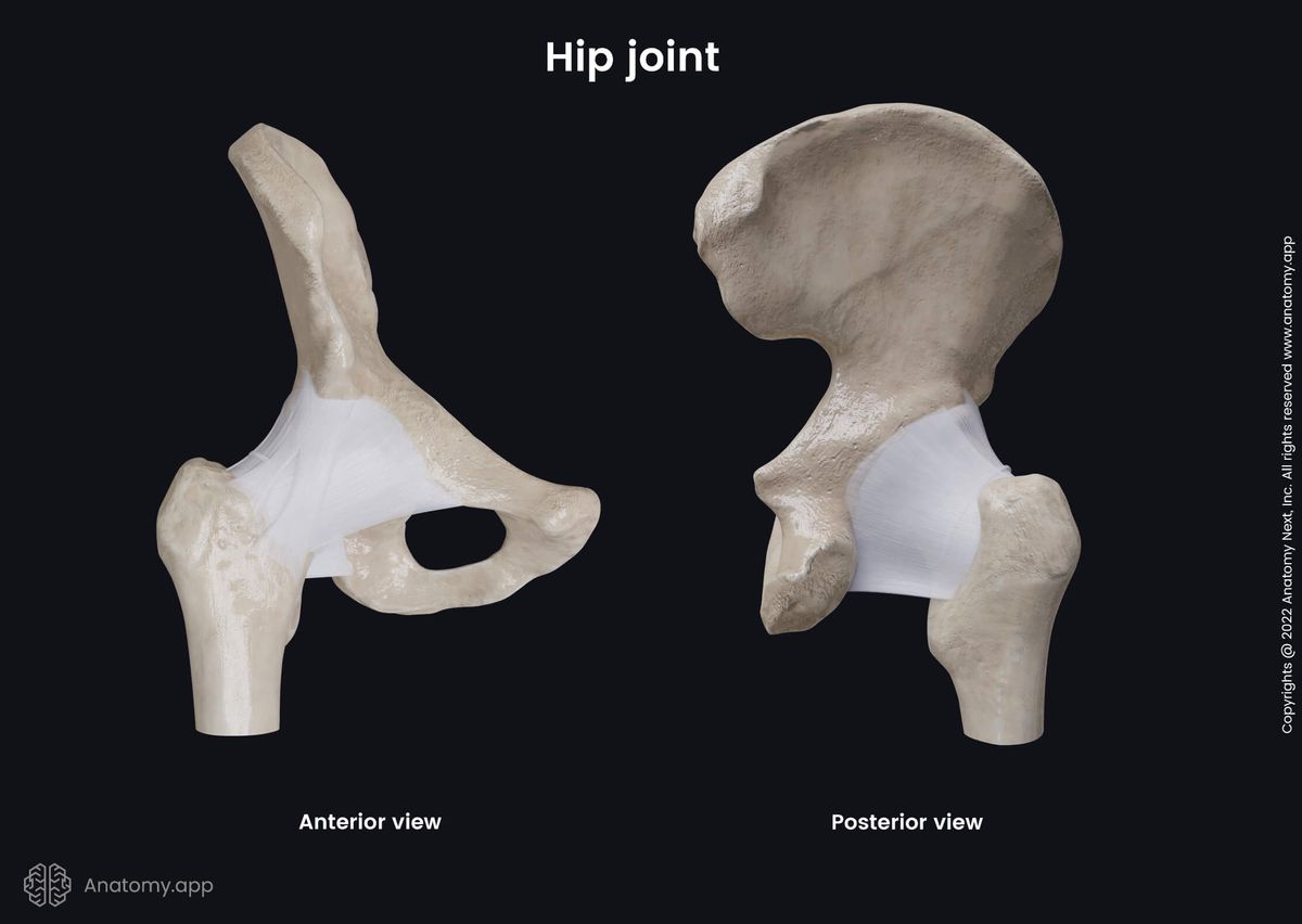
Hip joint
The hip joint (Latin: articulatio coxae), also known as the coxofemoral or acetabulofemoral joint, is an articulation formed between the acetabulum of the pelvis and the head of the femur. Therefore, the hip joint connects the pelvic girdle to the lower free extremity. It is classified as the synovial ball and socket type joint.
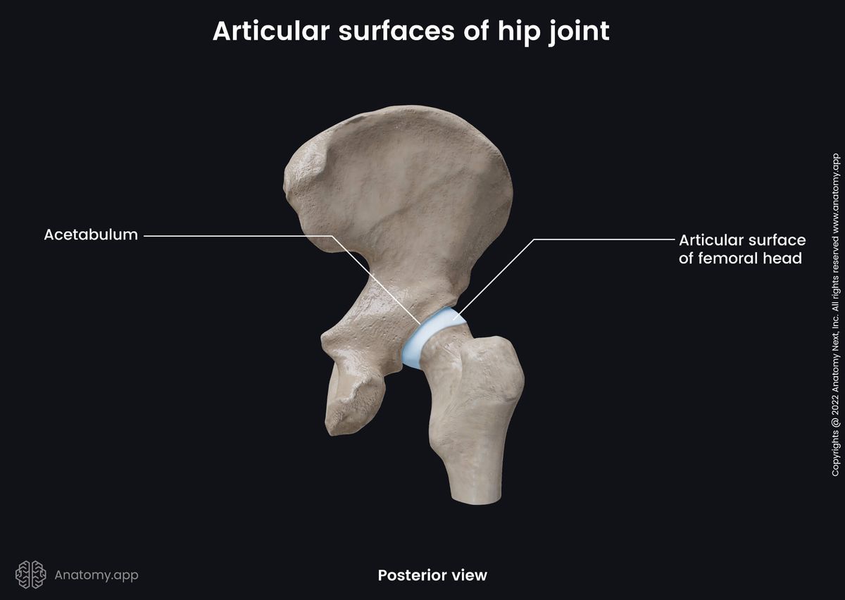
Articulating structures of hip joint
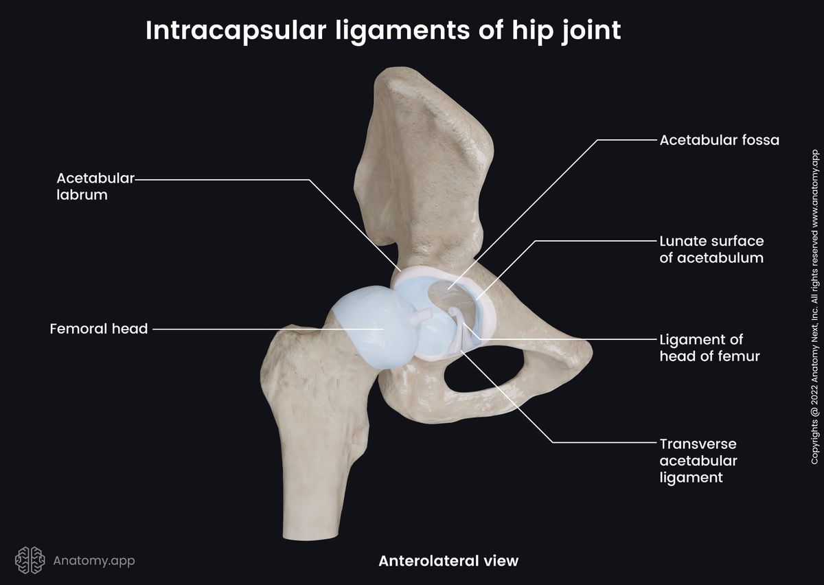
The articulating surfaces of the hip joint are as follows:
- Articular surface of the femoral head
- Lunate surface of the acetabulum
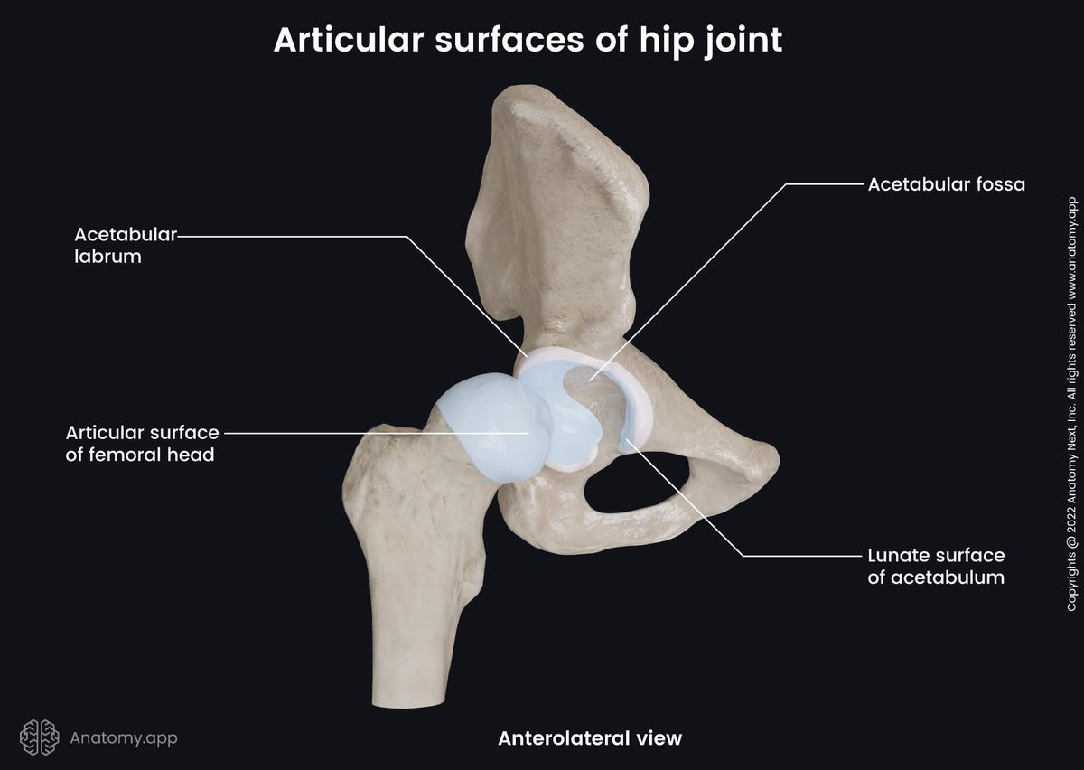
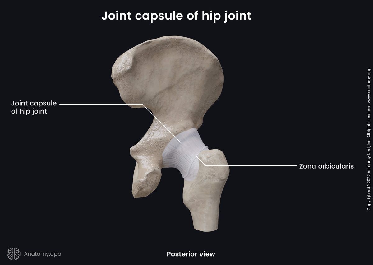
The acetabulum is a cup-like depression on the inferolateral aspect of the pelvis. It is surrounded by a fibrocartilaginous collar - the acetabular lip or acetabular labrum - that is attached to the external margins of the acetabulum. Both articulating structures are enclosed in a fibrous joint capsule that is attached to the outer edge of the acetabulum and acetabular labrum.
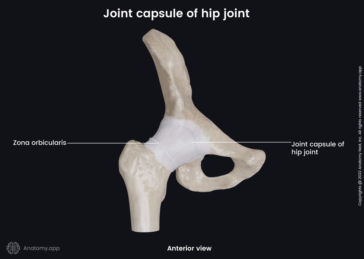
Ligaments of hip joint
The stability of the hip joint is increased with the help of two ligament groups. They are named based on their location in relation to the fibrous capsule - intracapsular and extracapsular ligaments.
The intracapsular ligaments are located inside the hip joint cavity. In contrast, the extracapsular ligaments are situated outside the capsule and are continuous with the outer surface of it.
Intracapsular ligaments
The hip joint cavity contains two intracapsular ligaments. They are known as follows:
- Transverse acetabular ligament - connects the ends of the lunate surface and passes above the acetabular notch;
- Ligament of the head of the femur - a small structure that runs from the acetabular notch and transverse acetabular ligament to the fovea capitis of the femoral head; through the ligament goes the artery to the head of the femur.
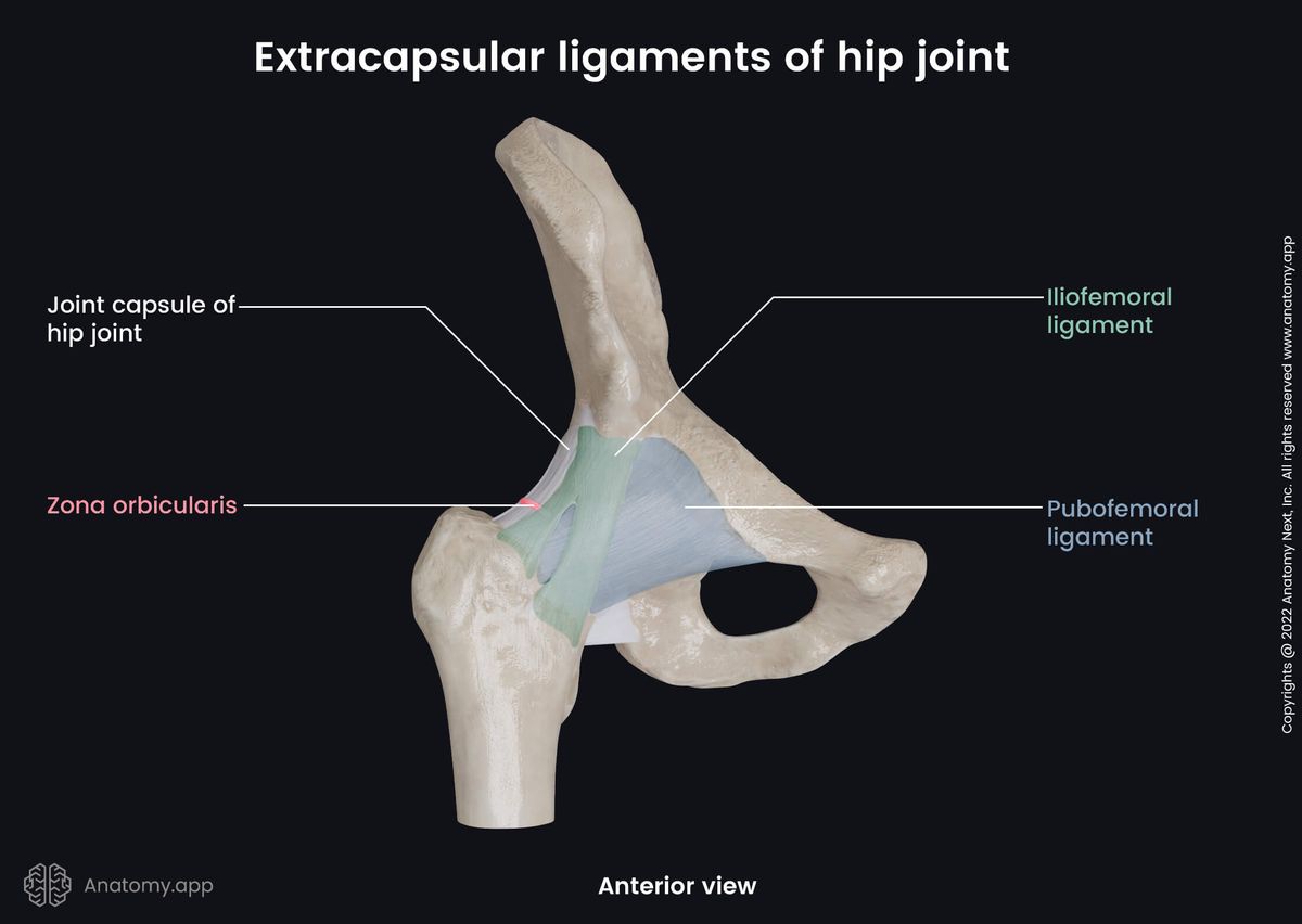
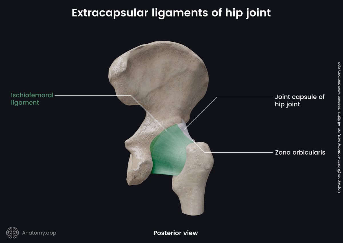
Extracapsular ligaments
The extracapsular ligaments include four strong bands. These ligaments are as follows:
- Iliofemoral ligament - extends between the anterior inferior iliac spine and the intertrochanteric line of the femur; it is the strongest ligament in the human body; it prevents external rotation and hyperextension of the hip joint;
- Pubofemoral ligament - triangular-shaped ligament located between the superior pubic ramus and the intertrochanteric line of the femur; it prevents the excessive abduction and inner rotation of the thigh at the hip joint;
- Zona orbicularis - extends from the anterior inferior iliac spine, surrounds the neck of the femur and ends where it started; it is formed by the circular fibers of the hip joint capsule;
- Ischiofemoral ligament - stretches from the body of the ischium to the trochanteric fossa; it prevents the thigh from excessive inner rotation and adduction at the hip joint.
Movements of hip joint
The hip joint is classified as a multiaxial joint, allowing a wide range of motions. However, the movements can be performed with a limited amplitude as this joint is the most important weight-bearing joint and it is more designed to provide stability to the body. The movements provided by the joint are as follows:
- Flexion and extension of the thigh
- Abduction and adduction of the thigh
- External or lateral rotation of the thigh
- Internal or medial rotation of the thigh
- Circumduction of the thigh
Contact information
- For questions regarding business inquiries. Please contact:
- info@anatomy.app
Product
Platform
- Terms & Conditions
- Privacy policy
- Imprint
- .
Anatomy Next © 2024. All rights reserved.