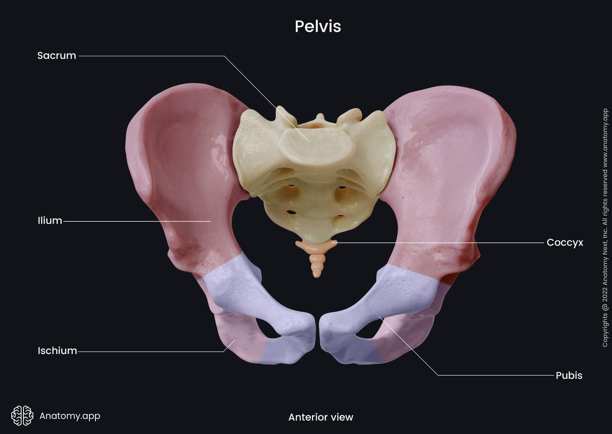
- Anatomical terminology
- Skeletal system
- Joints
- Muscles
- Heart
- Blood vessels
- Nervous system
- Respiratory system
- Digestive system
- Lymphatic system
- Female reproductive system
- Male reproductive system
- Endocrine glands
- Eye
- Ear
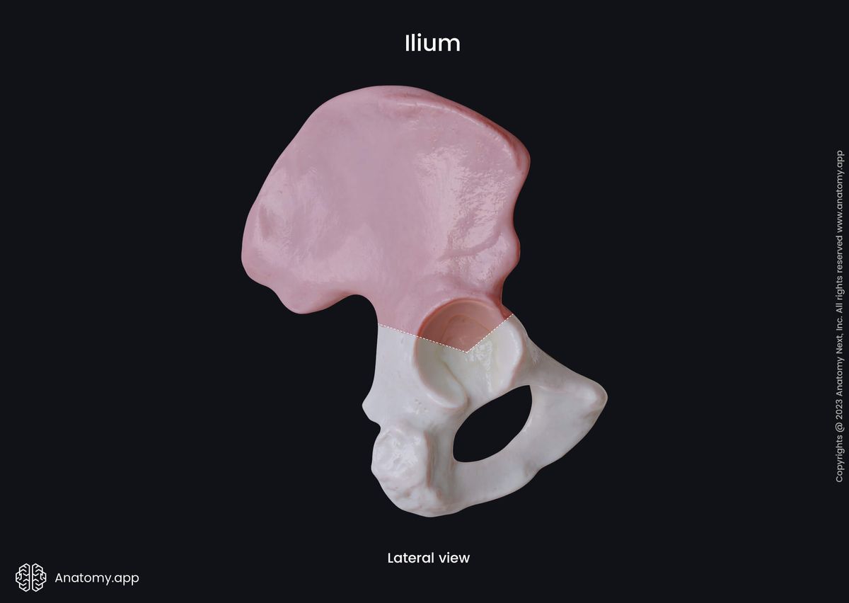
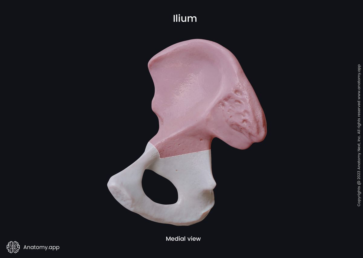
Ilium
The ilium (Latin: os ilium) is also known as the iliac bone. It is a paired bone forming the uppermost and most significant part of the hip bone and the skeleton of the pelvis.
Parts of ilium
Each iliac bone is composed of two parts, and they are as follows:
- Body
- Wing
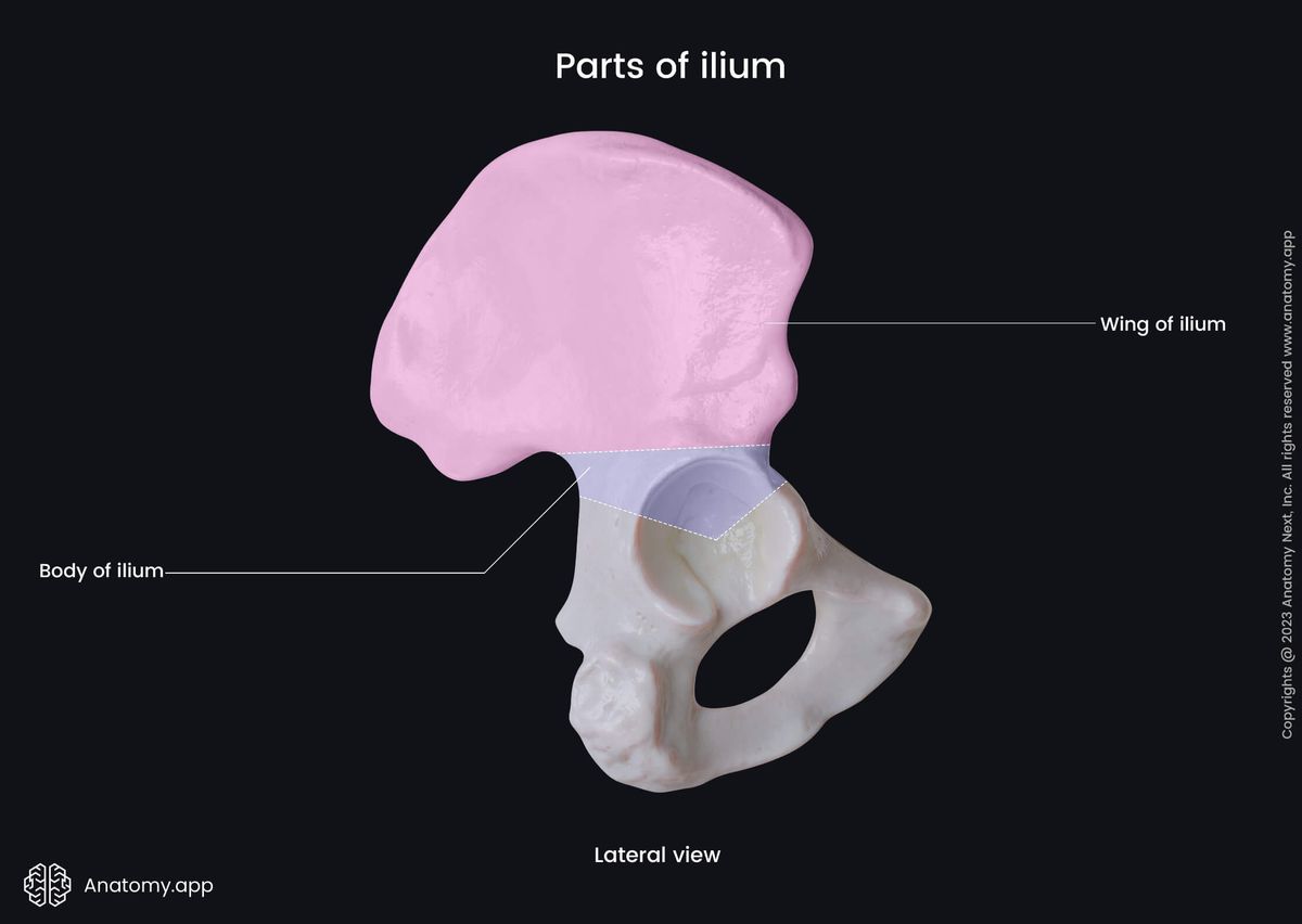
The body of the ilium is the central portion located above the acetabulum. Part of the body also contributes to forming one-third of the acetabulum.
The internal surface of the iliac body forms a part of the lesser pelvis. It also serves as the origin site for some fibers of the obturator internus muscle.
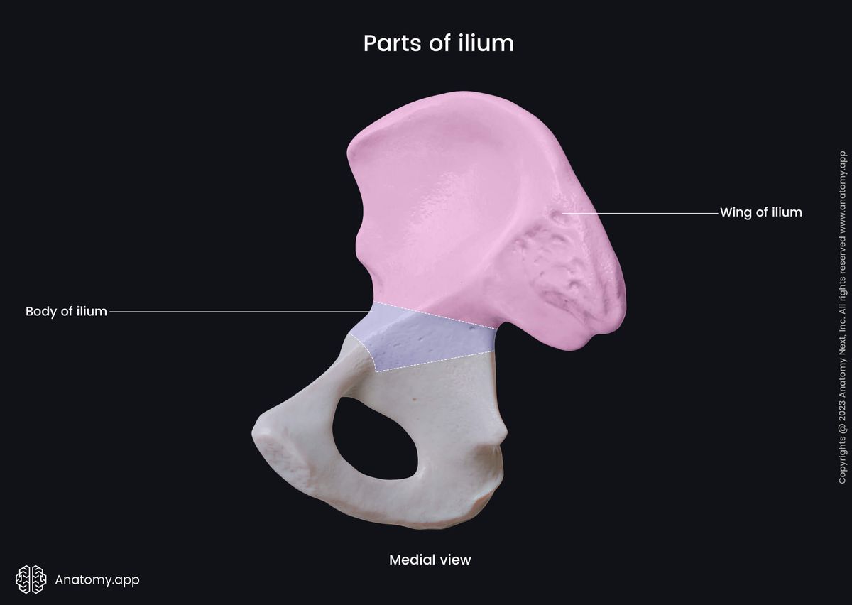
The wing of the ilium is the large expanded superior portion of the ilium. The wings of the ilium represent the lateral borders of the greater pelvis.
Wing of ilium
The wing of ilium, also known as the ala of the ilium, is the largest upper part of the bone. Therefore it contains several important landmarks, and these landmarks include the following:
- Arcuate line
- Iliac crest, featuring
- Anterior superior iliac spine
- Anterior inferior iliac spine
- Posterior superior iliac spine
- Posterior inferior iliac spine
- Iliac fossa
- Sacropelvic surface, presenting
- Auricular surface
- Iliac tuberosity
- Gluteal surface, featuring
- Anterior gluteal line
- Posterior gluteal line
- Inferior gluteal line
The arcuate line is a prominent bony ridge located on the internal surface of the ilium. It separates the body and the wing of the ilium and also forms the boundary between the greater and lesser pelvis.
The iliac crest is the superior border of the wing, and it also forms the superolateral margin of the greater pelvis. The iliac crest presents four bony projections - anterior et posterior superior and inferior iliac spines.
The anterior superior iliac spine is a bony prominence that marks the anterior limit of the iliac crest and is the most anterior part of the ilium. It also serves as the origin site for the sartorius muscle.
The anterior inferior iliac spine is a bony prominence located below the anterior superior iliac spine on the anterior aspect of the wing. It serves as an origin site for the rectus femoris muscle.
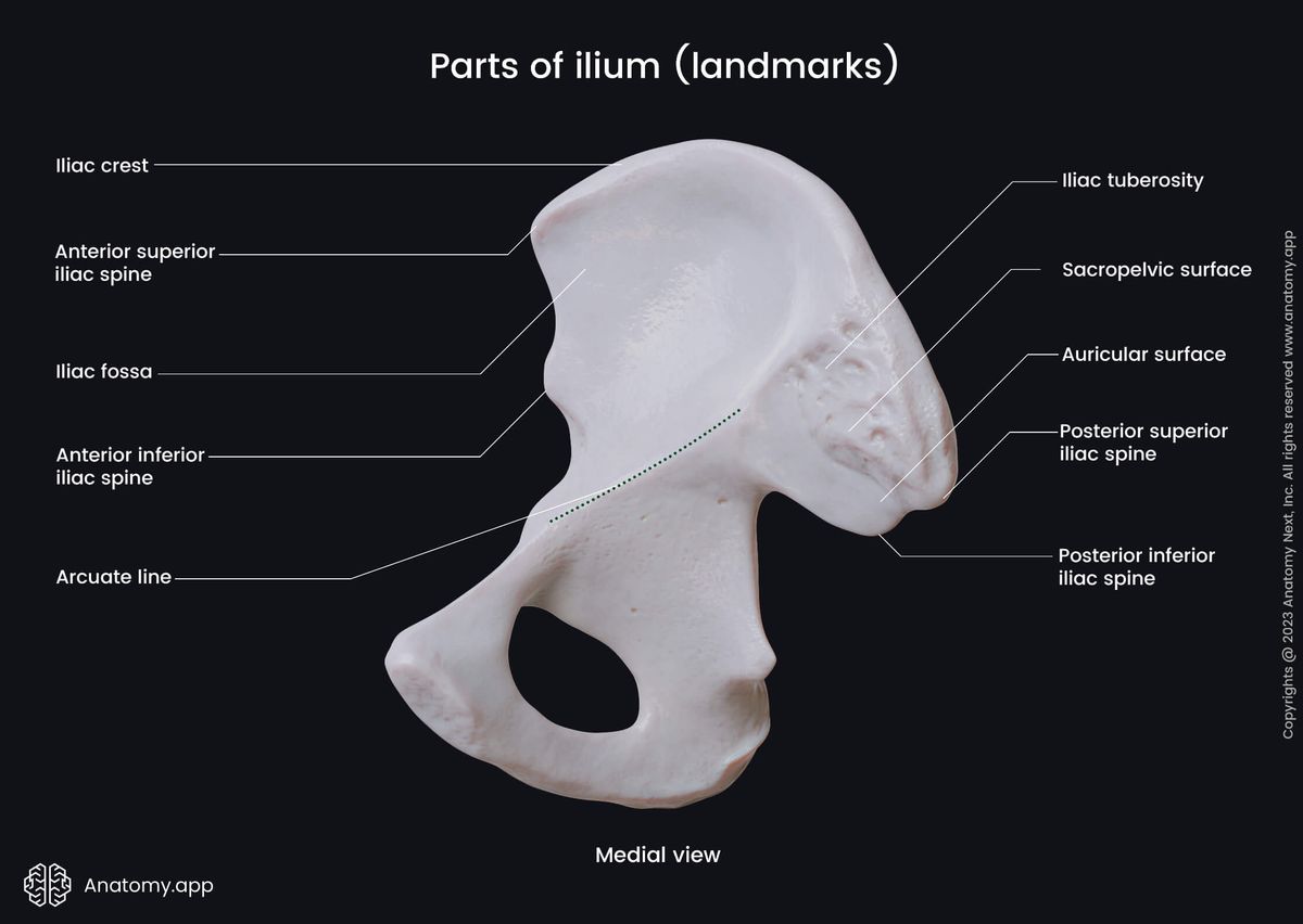
The posterior superior iliac spine is also a bony prominence marking the posterior limit of the iliac crest. It is an attachment site for the posterior sacroiliac ligament and the lumbar multifidus muscles.
The posterior inferior iliac spine is a bony prominence situated below the posterior superior iliac spine on the posterior aspect of the wing. Below this spine is located a deep notch called the greater sciatic notch.
The iliac fossa is a large and smooth concavity located on the internal surface of the wing. It is positioned on the anterior aspect of the surface. It serves as the origin site for the iliacus muscle.
The sacropelvic surface of the ilium corresponds to the posterior aspect of the inner surface. It is the part of the posterior portion that faces the sacrum. This surface consists of two parts - the auricular surface and the iliac tuberosity.
The auricular surface is an ear-shaped articular surface located within the sacropelvic surface. It articulates with the sacrum, and the joint between both bones is known as the sacroiliac joint. The auricular surface is covered with fibrocartilage.
The iliac tuberosity is a roughened and elevated area behind and above the auricular surface. It serves as an attachment site for the posterior sacroiliac ligaments and as an origin site for the lumbar multifidus muscles.
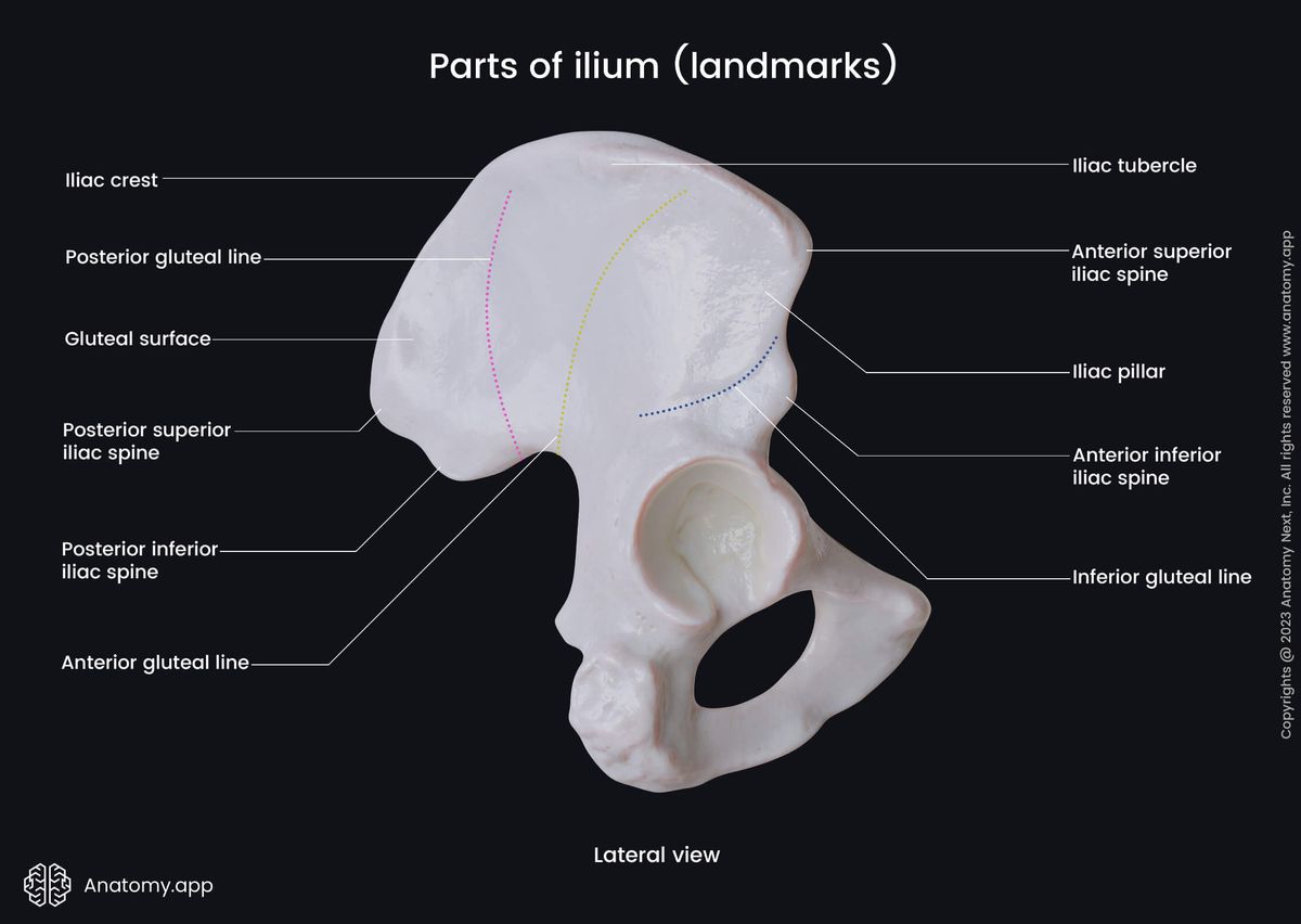
The gluteal surface of the ilium is the external surface of the wing. This surface presents with three ridges - the anterior, posterior and inferior gluteal lines.
The anterior gluteal line is a flat ridge situated almost in the middle of the wing on its external surface. It is located between the sites of origin of the gluteus medius and gluteus minimus muscles. The anterior gluteal line is the longest of all three lines.
The posterior gluteal line is also a bony ridge on the outer surface of the wing. It is located posterior to the anterior gluteal line and between the origin sites of the gluteus medius and gluteus maximus muscles.
The inferior gluteal line is a bony ridge located above the acetabulum and inferior to the anterior gluteal line. It is positioned between the origin sites of the gluteus minimus and rectus femoris muscles.
Anatomy.app
Contact information
- For questions regarding business inquiries. Please contact:
- info@anatomy.app