How to use Anatomy.app to study the cranial nerves
One of the most challenging topics when learning anatomy are the cranial nerves. All of these nerves have a specific function and they are in charge of many of the sensory and motor responsibilities of the human nervous system. This topic is even more difficult in medical school because there is little time to absorb all the information. The only way then to learn the nerves is to use an efficient tool that is comprehensive and capable of clearly conveying a large amount of information. One such tool that has all the mentioned qualities is Anatomy.app. In this article I will be discussing how to study the cranial nerves using Anatomy.app.
Go through the overview slide
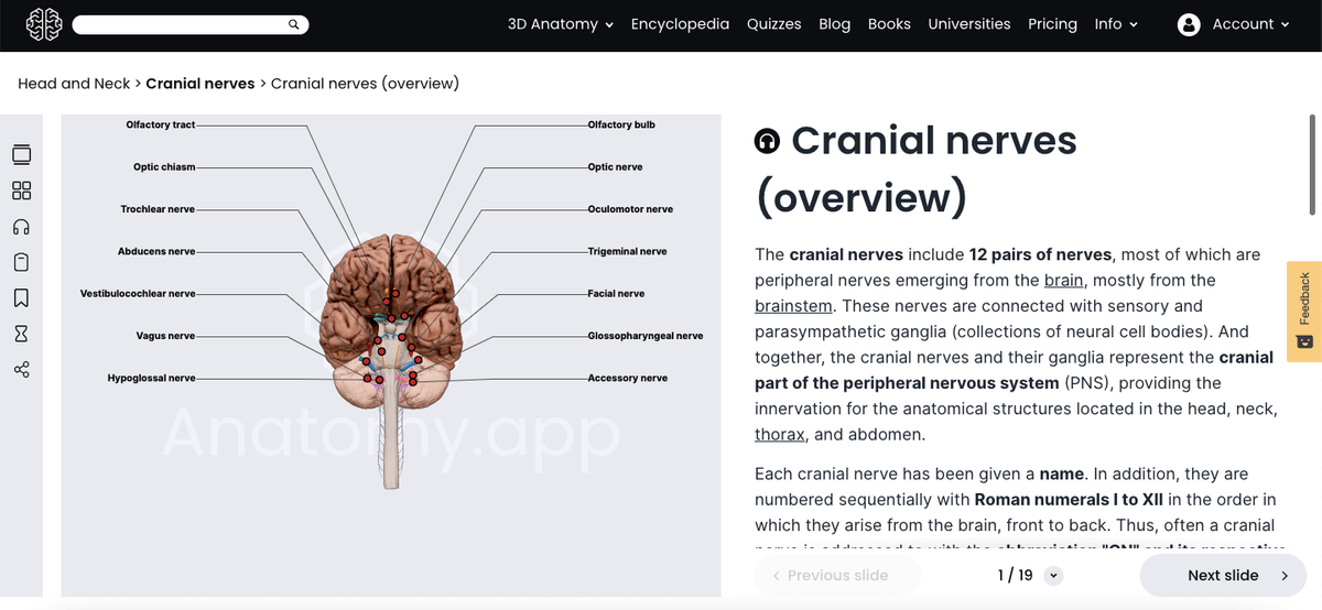
Go through the slides in the beginning just to get an overview of all the cranial nerves. You want to start getting your brain used to seeing where the cranial nerves are and what their general location is. When using the 3D image view of the brainstem, make sure you know where each nerve originates and at what level it arises (pons, medulla oblongata, or midbrain). Anatomy.app makes this easy to memorize because they have color-coded each nerve.
Additionally, the overview slide has a helpful mnemonic device to help you remember the nerves.
Draw or describe the pathway
Using the 3D anatomy, try to see if you can draw or describe the course of each nerve without looking at the screen. Anatomy.app makes it easy to follow the pathway of the nerves from multiple views, so you are able to see where the nerves enter and exit the skull. Once you have created a good visual image in your head of how the nerve goes, grab a buddy or a friend and see if you can describe its course to them. Sometimes I like to pretend like I am teaching them anatomy.
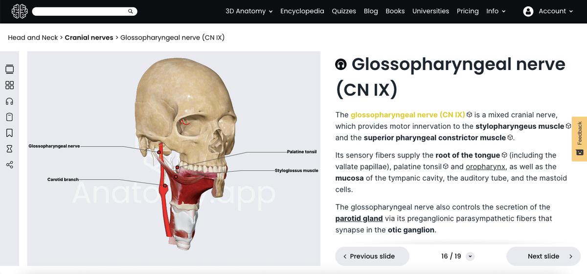
Use the toggle function
Use the toggle function to see the nerves from various viewpoints. For example, the cranial nerve IX (glossopharyngeal nerve) innervates the root of the tongue, parotid gland and the oropharynx. This is quite difficult and almost impossible to see from only one viewpoint.
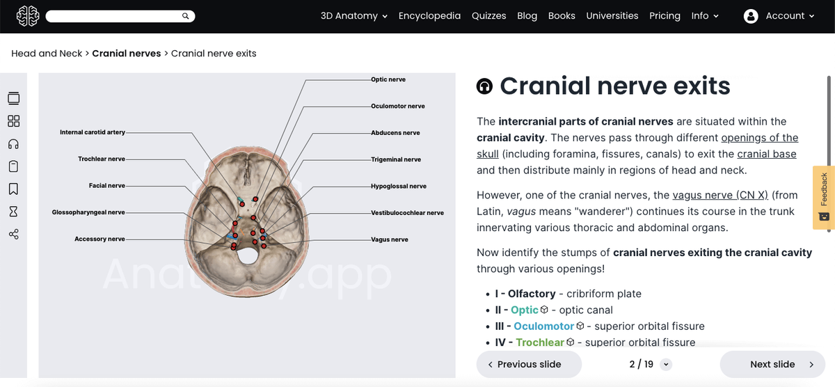
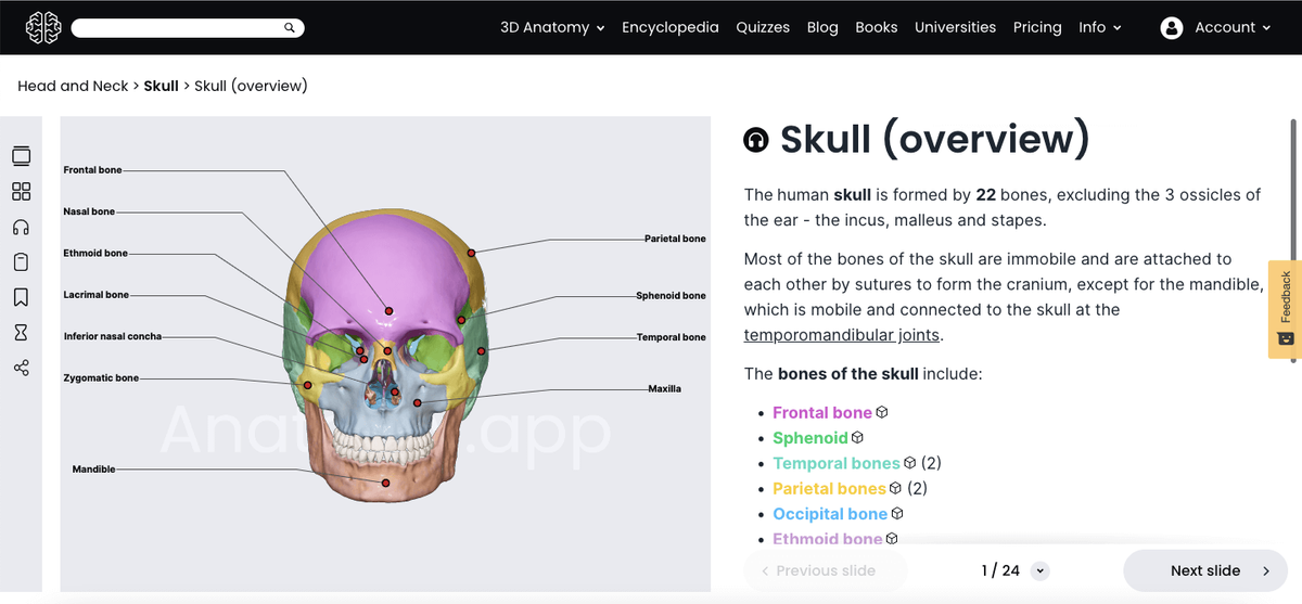
Learn what the skull looks like
The cranial nerves all have unique paths, and they originate from different levels of the brainstem. Therefore, the cranial nerves leave the skull through various openings. For example, the cranial nerves III, IV and VI all travel through the superior orbital fissure, while the cranial nerves IX, X and XI pass through the jugular foramen.
The thing that I love is that Anatomy.app contains detailed 3D articles about the skull, and also offers a comprehensive guide on the anatomy of the skull that can be purchased on the website. Alternatively, you can visit the Encyclopedia section, which is free of charge and goes into depth about the anatomy of the skull.
Recognize surrounding structures
Being able to recognize the surrounding structures can help you identify cranial nerves based on spatial cues. For example, the cranial nerves III and VI are easy to spot if you know where the midbrain or the pontomedullary junction is. Likewise, knowing where the internal carotid artery is can help you identify the carotid branch of the glossopharyngeal nerve.
Determine if the nerve is sensory, motor, or mixed
The type of the cranial nerves is a very common question that comes up in exams. The way I remember it is by thinking about all of the functions that the nerve provides. The slides in Anatomy.app make a note of this. One way you can memorize whether the cranial nerve is a motor, sensory or mixed nerve is by seeing which structures and muscles are near it.
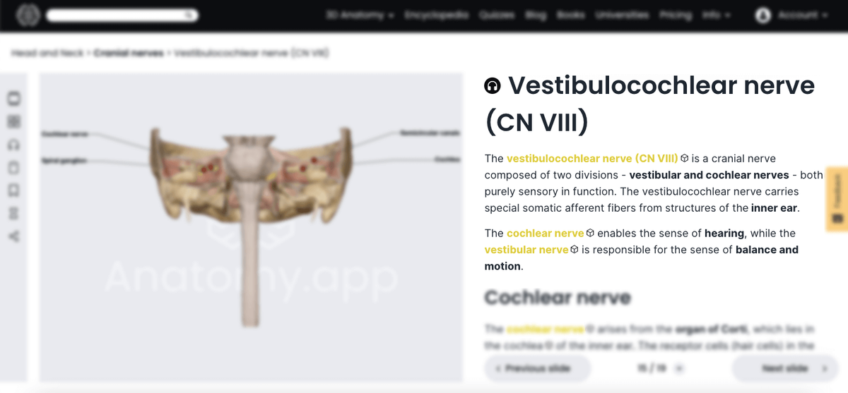
Try to imagine what would happen if one of the cranial nerves got damaged
Try to quiz yourself. For example, you might ask yourself something like what does a person with facial nerve palsy look like? Or what happens when the vestibulocochlear nerve is damaged? Anatomy.app helps with this because it puts the functions of the cranial nerves in bold.
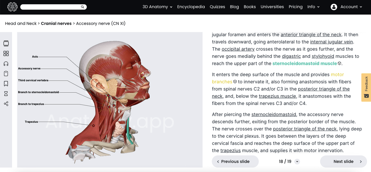
Try to locate where the nerve would be if it were covered with other tissue
You can try to locate where the nerve would be if more layers of tissue were situated above it. Try to see which muscles the nerve passes through and which muscle groups are near the nerve but are not necessarily innervated by it. And again, Anatomy.app makes it easy because one of its 3D images is a head that is dissected down to the level of muscles.
Quiz yourself
The best way to master the content is to quiz yourself. Fortunately, the interface on Anatomy.app allows you to take quizzes on every article found on the platform, and the cool thing is that you can also choose the difficulty level of the quiz. To access the quiz, you just have to go to the top right-hand corner of the website, where you can select the option quizzes, and here you go - you are ready to dare yourself.

I suggest first mastering all of the questions in the Anatomy.app question bank, then further supplement your learning with other videos.
Learn one nerve at a time
It is important to really take your time and ensure you have a sound understanding. Breaking up large amounts of information into smaller components has been shown to be a more effective learning method than just consuming all of the information at once.
The cranial nerves are definitely one of the most complicated topics in anatomy. Fortunately, there are websites such as Anatomy.app that have everything you need to not only pass but excel in your anatomy class.
Written by
Eric Mubang
M.D. candidate
http://fitmedicwriter.com/
Anatomy.app
Contact information
- For questions regarding business inquiries. Please contact:
- info@anatomy.app