- Anatomical terminology
- Skeletal system
- Joints
- Muscles
- Heart
- Blood vessels
- Blood vessels of systemic circulation
- Aorta
- Blood vessels of head and neck
- Blood vessels of upper limb
- Blood vessels of thorax
- Blood vessels of abdomen
- Blood vessels of pelvis and lower limb
- Blood vessels of systemic circulation
- Nervous system
- Respiratory system
- Digestive system
- Lymphatic system
- Female reproductive system
- Male reproductive system
- Endocrine glands
- Eye
- Ear
Aorta
The aorta (Latin: aorta) is the largest blood vessel in the human body and the main artery of systemic circulation. It transports oxygenated blood from the heart to the rest of the body. It arises from the left ventricle of the heart, ascends for a short distance, forms an arch and further travels down to the abdomen. The aorta is around one foot (30 cm) long and has complex hemodynamic functions. It is typically divided into the following four parts:
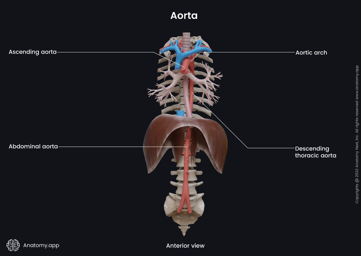
The first three parts are located within the thorax, and therefore, they are considered portions of the thoracic aorta. The rest of the aorta is situated in the abdominal cavity and is called the abdominal aorta, respectively.
The thoracic aorta originates at the level of the aortic valve of the heart. It continues as the abdominal aorta when it pierces the diaphragm and enters the abdominal cavity via the aortic hiatus. It happens at the level of the twelfth thoracic vertebra (T12).
Overall, the thoracic aorta is divided into ascending, transverse and descending portions. The ascending part is referred to as the ascending aorta, while the transverse part forms an arch and therefore is known as the aortic arch. The descending part is usually called the thoracic aorta.
However, the descending part of the aorta may be referred to as both - the thoracic and abdominal parts - as they both go down toward the pelvis. Therefore, the descending part of the thoracic aorta is usually called the descending thoracic aorta for clarity and understanding, while the abdominal part is simply called the abdominal aorta.
Ascending aorta
The ascending aorta (Latin: aorta ascendens) is the first part of the aorta. It is typically only around 2 inches (5 cm) long. However, it is the widest part of the aorta, and its diameter is about 1.3 inches (3 cm). It continues as the aortic arch at the upper border of the second right costal cartilage.
The ascending aorta originates from the aortic orifice at the base of the left ventricle, approximately at the inferior border of the third left costal cartilage behind the left half of the sternal body.
The aortic orifice is guarded by the aortic valve that separates the ascending aorta from the left ventricle. From the origin site, the ascending aorta goes obliquely upward, anterior and slightly to the right side.
The ascending aorta is composed of two parts:
- The root of the aorta is the first portion that begins from the annulus of the aortic valve and extends to the sinotubular junction. The aortic annulus is a fibrous skeletal frame to which are attached the aortic valve cusps. The aortic root includes the aortic sinuses (sinuses of Valsalva), which are three small anatomic dilatations found above the aortic valve. Overall, the root of the aorta appears enlarged with a characteristic bulge.
- The tubular ascending aorta is the second portion that extends from the sinotubular junction to the aortic arch. This portion usually ends at the level of the first branch of the aortic arch - the brachiocephalic trunk.

The ascending aorta gives off only two branches, and it happens at the coronary sinuses level. The right coronary sinus is an origin site for the right coronary artery (RCA), while the left coronary sinus gives rise to the left coronary artery (LCA). They supply the myocardium (heart muscle) and essential heart structures with arterial blood.
The ascending aorta is one of the great vessels of the heart and is located within the pericardium. Therefore, it is considered the content of the middle compartment (middle mediastinum) of the inferior mediastinum.
Related structures of ascending aorta
Superiorly, the ascending aorta is related to the pleura, anterior border of the right lung, thymus remnant and sternum. Anteriorly, it connects with the conus arteriosus, right auricle and initial segment of the pulmonary trunk.
Posteriorly, the ascending aorta is related to the left atrium, right main (principal) bronchus and right pulmonary artery. And finally, the right atrium and superior vena cava are situated laterally on the right side, while the left atrium and pulmonary trunk lie laterally on the left side.
Aortic arch
The aortic arch (Latin: arcus aortae) is the transverse part of the thoracic aorta found between its ascending and descending portions. It begins posterior to the right second sternocostal joint, as the ascending aorta emerges from the pericardium. Overall, the aortic arch provides arterial blood supply to the head, neck and upper limbs.
The aortic arch is positioned within the superior mediastinum of the thorax. From its origin, it passes superiorly, posteriorly and to the left before it descends. The aortic arch ends on the left side at the level of the fourth thoracic vertebrae (T4).
At first, it lies anterior to the tracheal bifurcation, but as it forms an arch, it is found on the left side of the trachea and esophagus. The aortic arch continues as the descending thoracic aorta at the level of the fourth thoracic vertebra (T4) or left second sternocostal joint.
Initially, the diameter of the aortic arch is larger and matches the ascending aorta. Gradually its diameter becomes smaller. At the site where it transitions into the descending aorta, the aortic arch has a small stricture called the aortic isthmus.
The aortic isthmus marks the border between the origin of the left subclavian artery and the connection of the ductus arteriosus to the descending thoracic aorta. After the aortic isthmus, the aorta becomes more dilated again.
Branches of aortic arch
The aortic arch gives off three branches that provide arterial blood supply to the head, neck and upper limbs. They include the following arteries:
- Brachiocephalic trunk
- Left common carotid artery
- Left subclavian artery
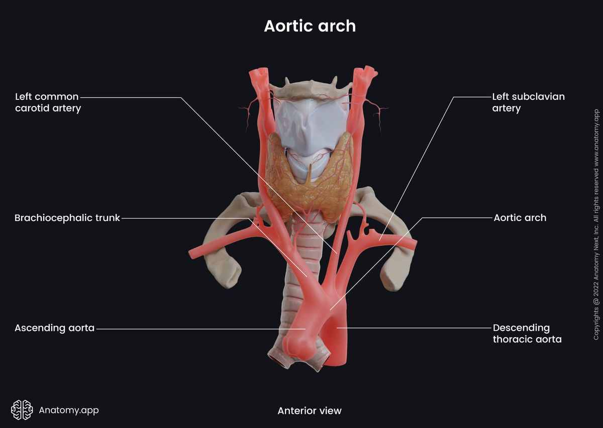
The brachiocephalic trunk is the first and largest branch of the aortic arch. It is also known as the brachiocephalic or innominate artery. It is a short vessel located within the superior mediastinum, right behind the manubrium of the sternum. The brachiocephalic trunk extends superiorly and posteriorly to the right side and marks the junction between the aortic arch and ascending aorta. Overall, it supplies arterial blood to the right side of the head, neck and right upper limb.
The brachiocephalic trunk terminates at the superior margin of the right sternoclavicular joint by dividing into two branches - the right common carotid artery and the right subclavian artery. The right common carotid artery supplies blood to the right side of the neck and head, including the right side of the brain. In contrast, the right subclavian artery supplies the right upper extremity, as well as it provides arterial blood supply to the head via the right vertebral artery.
The left common carotid artery is the second and, at the same time, the longest branch of the aortic arch. Overall, it supplies the head and neck on the left side. After it arises, it ascends and exits the thorax via the superior thoracic aperture. At this point, it is located behind the left sternoclavicular joint. In the neck, it crosses the carotid triangle and ascends within the carotid sheath. At the level of the superior border of the thyroid cartilage, the left common carotid artery divides into two terminal branches - the left external carotid artery and the left internal carotid artery.
The left subclavian artery is the smallest branch of the aortic arch. It supplies arterial blood to the head, brain, neck, upper thorax, left shoulder and upper limb. Like the left common carotid artery, the left subclavian artery exits the thorax via the superior thoracic aperture. At first, it passes between the anterior scalene muscle and the sternal end of the clavicle, and then it goes between the anterior and middle scalene muscles. And finally, the left subclavian artery travels between the first rib and clavicle as a part of a neurovascular bundle consisting of the subclavian artery, subclavian vein and brachial plexus. At the outer border of the first rib, the left subclavian artery becomes the left axillary artery.
Related structures of aortic arch
Superiorly, the aortic arch is related to the brachiocephalic trunk, left brachiocephalic vein, left common carotid artery and left subclavian artery. Also, the thymus remnant is located superior to it.
Inferiorly, the arch relates to the pulmonary trunk and its bifurcation, left main (principal) bronchus, left recurrent laryngeal nerve, superficial cardiac plexus and ligamentum arteriosum. As the left vagus nerve (CN X) travels across the aortic arch, it gives off the recurrent laryngeal nerve, which curves around the arch and ascends back towards the neck.
Anteriorly and more to the left side lies the left lung and mediastinal pleura. Also, the aortic arch is crossed by the following structures - left phrenic nerve, left inferior cardiac branch of the vagus nerve (CN X), left superior cervical cardiac branch of the sympathetic trunk, left superior intercostal vein and left vagus nerve. Anterior to the aortic arch lies the thymus remnant.
Posteriorly and more to the right side are located the trachea, esophagus, left recurrent laryngeal nerve, deep cardiac plexus and thoracic duct. Also, posterior to the aortic arch is the spine.
Descending thoracic aorta
The descending thoracic aorta (Latin: aorta descendens thoracica) is the longest portion of the aorta and a continuation of the aortic arch. It is positioned in the posterior compartment of the inferior mediastinum. The descending thoracic aorta starts at the left side of the body of the fourth thoracic vertebra (T4).
The proximal portion of the descending thoracic aorta typically has a slight outpouching called the ductus diverticulum (ductus bump). This bulge is found on the anterior and medial aspects of the descending thoracic aorta, representing a remnant of ductus arteriosus that functions during the embryonic period.
As it descends along the bodies of the fifth to twelfth thoracic vertebrae (T5 - T12), the descending thoracic aorta moves from the left side of the spine towards its midline. When reaching the aortic hiatus of the diaphragm, it is positioned anterior to the body of the twelfth thoracic vertebra (T12). As the descending thoracic aorta passes through the diaphragm, it continues as the abdominal aorta.
Branches of descending thoracic aorta
The descending thoracic aorta gives off various branches that can be subdivided into two groups based on the structures they supply. The visceral branches supply the structures and internal organs found within the mediastinum, such as the pericardium, bronchi and esophagus. In contrast, the parietal branches of the descending thoracic aorta provide arterial supply to the walls of the thorax.
Visceral branches of descending thoracic aorta
The visceral branches of the descending thoracic aorta include the following arteries:
- Pericardial branches - supply the posterior and lateral aspects of the pericardium;
- Bronchial branches - variable in size and number; two left bronchial arteries (superior and inferior) directly arise from the descending thoracic artery, while the right lung has only one bronchial artery with a variable origin; it usually arises from the intercostobronchial trunk; they supply the roots of the lungs, visceral pleura, thoracic esophagus, pulmonary vessels, trachea, bronchi, connective tissue of the lungs and pericardium;
- Esophageal branches - up to five vessels that supply the thoracic portion of the esophagus;
- Mediastinal branches - supply the structures of the posterior mediastinum, including lymph nodes, nerves, blood vessels, areolar tissue, adipose tissue and other structures.
Parietal branches of descending thoracic aorta
The parietal branches of the descending thoracic aorta are as follows:
- Superior phrenic branches - originate from the distal part of the descending thoracic aorta; they distribute within the thoracic surface of the diaphragm, supplying its posterior aspect; these arteries anastomose with the musculophrenic and pericardiacophrenic arteries;
- Posterior intercostal arteries - nine pairs of arteries that arise from the posterior aspect of the descending thoracic aorta and supply the third to eleventh intercostal spaces; anteriorly, these arteries anastomose with either the internal thoracic or musculophrenic arteries;
- Subcostal arteries - a pair of arteries that travel within the subcostal space found below the last rib; these arteries mainly supply the flat abdominal muscle, such as the external abdominal oblique, internal abdominal oblique and transversus abdominis muscles.
Related structures of descending thoracic aorta
Anteriorly, the descending thoracic aorta borders with the left main (principal) bronchus, hilum of the left lung, pericardium, left atrium, lower portion of the esophagus and diaphragm. Posteriorly to it lies the thoracic spine, hemiazygos vein, accessory hemiazygos vein and sympathetic trunk.
Laterally to the right are the thoracic duct, superior portion of the esophagus, azygos vein, right lung and mediastinal pleura. Laterally to the left lie the left lung and mediastinal pleura.
Abdominal aorta
The abdominal aorta (Latin: aorta abdominalis) is a direct continuation of the descending thoracic aorta. It begins once the descending thoracic aorta reaches the abdominal cavity via the aortic hiatus of the diaphragm. It happens at the level of the inferior border of the twelfth thoracic vertebra (T12). The abdominal aorta descends within the retroperitoneal space anterior to the bodies of the lumbar vertebrae.
The abdominal aorta terminates at the level of the fourth lumbar vertebra (L4) (or the intervertebral disc between the fourth and fifth lumbar vertebrae (L4/L5)) by dividing into two large branches - the right and left common iliac arteries.
Branches of abdominal aorta
Like the branches of the descending thoracic aorta, the branches of the abdominal aorta can also be grouped into the visceral and parietal branches. Alternatively, they can be subdivided based on the side of the abdominal aorta from which they arise - anterior, lateral and posterior branches.
The anterior and lateral branches supply the internal organs, and all are visceral branches. In contrast, the posterior branches supply the abdominal walls, spine and contents of the vertebral canal. Therefore, the posterior branches are also the parietal branches.
Anterior branches of abdominal aorta
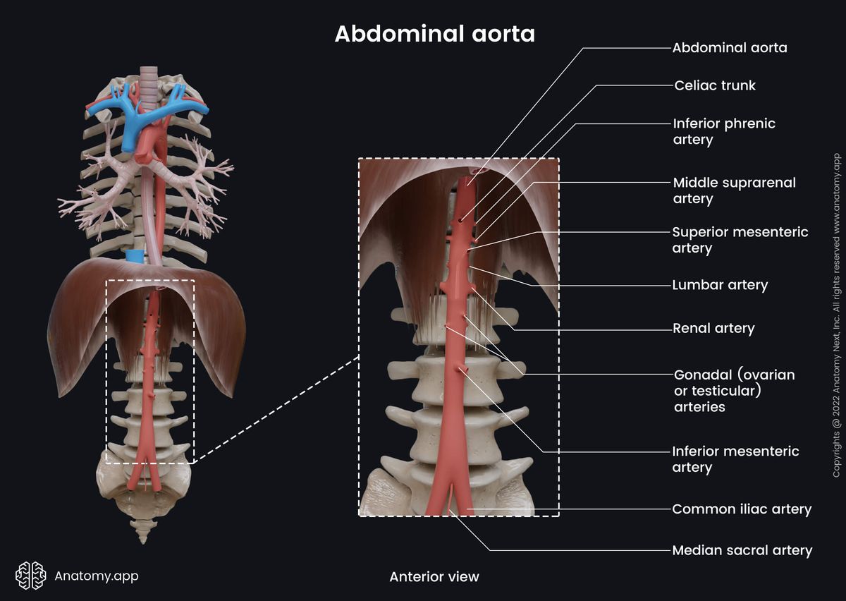
The anterior branches of the abdominal aorta are all unpaired arteries, and they are as follows:
- Celiac trunk
- Superior mesenteric artery
- Inferior mesenteric artery
The celiac trunk is the first branch of the abdominal aorta. It is a short trunk that arises from the anterior aspect of the abdominal aorta just below the aortic hiatus. It usually originates at the level of the inferior border of the body of the twelfth thoracic vertebra (T12). The celiac trunk immediately divides into three branches - the left gastric, common hepatic and splenic arteries. Overall, the celiac trunk supplies arterial blood to the abdominal part of the esophagus, stomach, first part of the duodenum, pancreas, liver, gallbladder and spleen.
The superior mesenteric artery arises below the celiac trunk at the level of the first lumbar vertebra (L1). It travels inferiorly and anteriorly, entering the mesentery of the small intestine. Overall, the superior mesenteric artery supplies the midgut derivatives, which include the small intestine (except the first portion of the duodenum) and large intestine up to the proximal two-thirds of the transverse colon.
The inferior mesenteric artery originates from the anterolateral aspect of the abdominal aorta at the level of the third lumbar vertebra (L3). Overall, it supplies the hindgut derivatives - the distal one-third of the transverse colon, descending colon, sigmoid colon, upper rectum and anal canal.
Note: During the embryological period, the gastrointestinal tract organs are derived from the primitive gut tube, which is divided into three portions: foregut, midgut and hindgut. Each of the portions is distinct in its embryological development and neurovascular supply. The foregut later develops into the esophagus, stomach, liver, gallbladder, pancreas, and the first part of the duodenum. The midgut develops into the rest of the duodenum, small intestine, and large intestine up to the proximal two-thirds of the transverse colon. And finally, the hindgut is the derivative of the remaining part of the large intestine up to the upper anal canal.
Lateral branches of abdominal aorta
The lateral branches of the abdominal aorta are paired arteries, and they include the following:
- Middle suprarenal arteries
- Renal arteries
- Gonadal arteries
The middle suprarenal arteries are two arteries arising from each side of the abdominal aorta near the superior mesenteric artery at the level of the first lumbar vertebra (L1). Each artery supplies the adrenal gland on its respective side and anastomoses with the suprarenal branches of the inferior phrenic and renal arteries.
The renal arteries are also a pair of arteries originating at the right angle from the lateral aspects of the abdominal aorta at the level of the first or second lumbar vertebrae (L1/L2). They supply the kidneys and give branches to the urinary bladder and adrenal glands.
The gonadal arteries include the ovarian arteries in females and testicular arteries in males. In either case, they are two long arteries arising from the lateral sides of the abdominal aorta below the origin site of the renal arteries at the level of the second or third lumbar vertebrae (L2/L3). Each artery supplies the ovary or testicle.
Posterior branches of abdominal aorta
The posterior branches of the abdominal aorta include the following vessels:
- Inferior phrenic arteries
- Lumbar arteries
- Median sacral artery
The inferior phrenic arteries are paired vessels that usually arise from the posterolateral aspect of the aorta superior to the celiac trunk at the level of the inferior border of the twelfth thoracic vertebra (T12). These arteries supply the abdominal surface of the diaphragm and give off several superior suprarenal arteries to the adrenal glands.
Each person usually has four pairs of lumbar arteries. They originate from the posterolateral aspects of the abdominal aorta from the first through fourth lumbar vertebrae (L1 - L4). These arteries supply the posterior and lateral abdominal walls. Also, they give branches to the deep back muscles and spinal cord.
The median sacral artery is a small artery that arises from the posterior surface of the abdominal aorta at the level of the fourth lumbar vertebra (L4) just above its bifurcation. It supplies the lower two lumbar vertebrae (L4 - L5), sacrum, coccyx, and it also gives some small branches to the posterior aspect of the upper rectum.
Terminal branches of abdominal aorta
The abdominal aorta bifurcates at the fourth lumbar vertebra (L4) level, dividing into two terminal branches - the right and left common iliac arteries. Overall, the common iliac arteries supply the pelvic and gluteal regions and the lower limbs.
Each common iliac artery travels posterolaterally along the medial border of the psoas major muscle. It terminates at the level of the sacroiliac joint by dividing into two terminal branches - the internal and external iliac arteries.
Related structures of abdominal aorta
Anteriorly, the abdominal aorta relates to the organs and blood vessels of the abdominal cavity, including the body of the pancreas, splenic vein and left renal vein, horizontal portion of the duodenum and liver.
Posteriorly to the aorta are the following structures - the twelfth thoracic vertebra (T12), first through fourth lumbar vertebrae (L1 - L4), their respective intervertebral discs and anterior longitudinal ligament.
Laterally on the right side of the abdominal aorta are the thoracic duct, cisterna chyli, azygos vein, inferior vena cava and right crus of the diaphragm.
Laterally on the left side of the abdominal aorta lies the left crus of the diaphragm. Also, the abdominal aorta is related to the ascending part of the duodenum and left sympathetic trunk.
References:
- Abbara, S., & Boxt, L. M. (2016). Cardiac imaging: The requisites. Elsevier.
- Gray, H., & Carter, H. (2021). Gray’s Anatomy (Leatherbound Classics) (Leatherbound Classic Collection) by F.R.S. Henry Gray (2011) Leather Bound (2010th Edition). Barnes & Noble.
- Kaufman, J.A., Lee, M.J. (2004). Vascular and Interventional Radiology. Elsevier.
- Moore, K.L., Dalley, A.F., Agur, A.M. (2018). Clinically Oriented Anatomy, 8th Edition, Lippincott Williams & Wilkins.
- Ţintoiu, I. C., Ursulescu, A., Elefteriades, J. A., Underwood, M. J., & Droc, I. (2018). New approaches to aortic diseases from valve to abdominal bifurcation. Academic Press.
- Townsend, C. M., Beauchamp, R. D., Evers, B. M., Mattox, K. L., & Christopher, F. (2022). Sabiston Textbook of Surgery: The Biological Basis of modern surgical practice. Elsevier.
Anatomy.app
Contact information
- For questions regarding business inquiries. Please contact:
- info@anatomy.app