- Head and Neck
- Dental Anatomy
- Upper Extremity
- Thorax
- Abdomen
- Spine and Back
- Pelvis
- Lower Extremity
- Organ Systems
- Anatomical terminology
- Skeletal system
- Skull
- Neurocranium
- Frontal bone
- Sphenoid bone
- Temporal bone
- Ethmoid bone
- Occipital bone
- Parietal bone
- Viscerocranium
- Lacrimal bone
- Nasal bone
- Zygomatic bone
- Palatine bone
- Maxilla
- Inferior nasal concha
- Vomer
- Mandible
- Hyoid bone
- Auditory ossicles
- Sutures of skull
- Topography of skull
- Cranial base
- Internal cranial base
- Anterior cranial fossa
- Middle cranial fossa
- Posterior cranial fossa
- External cranial base
- Temporal fossa
- Infratemporal fossa
- Pterygopalatine fossa
- Orbit
- Nasal cavity
- Paranasal sinuses
- Maxillary sinus
- Frontal sinus
- Sphenoidal sinus
- Ethmoidal air cells
- Skeleton of trunk
- Rib cage
- Ribs
- Sternum
- Thoracic vertebrae
- Spine
- Cervical vertebrae
- Thoracic vertebrae
- Lumbar vertebrae
- Sacrum
- Coccyx
- Skeleton of upper limb
- Bones of shoulder girdle
- Clavicle
- Scapula
- Humerus
- Bones of forearm
- Radius
- Ulna
- Bones of hand
- Carpal bones
- Metacarpal bones
- Phalanges of hand
- Skeleton of lower limb
- Pelvis
- Hip bone
- Pubic bone
- Ischium
- Ilium
- Sacrum
- Coccyx
- Femur
- Patella
- Bones of leg
- Tibia
- Fibula
- Bones of foot
- Tarsal bones
- Talus
- Calcaneus
- Cuboid bone
- Navicular bone
- Cuneiform bones
- Metatarsal bones
- Phalanges of foot
- Joints
- Classification of joints
- Joints of skull
- Temporomandibular joint
- Sutures of skull
- Joints of spine
- Anterior longitudinal ligament
- Posterior longitudinal ligament
- Supraspinous and nuchal ligaments
- Ligamenta flava
- Intervertebral discs
- Atlanto-occipital joint
- Atlanto-axial joint
- Facet (zygapophyseal) joints
- Lumbosacral joint
- Sacrococcygeal joint
- Joints of lower limb
- Joints of pelvis
- Sacrotuberous ligament
- Sacrospinous ligament
- Obturator membrane
- Pubic symphysis
- Sacroiliac joint
- Hip joint
- Knee joint
- Tibiofibular joints
- Superior tibiofibular joint
- Inferior tibiofibular joint
- Joints of foot
- Ankle joint
- Intertarsal joints
- Talocalcaneonavicular joint
- Calcaneocuboid joint
- Transverse tarsal joint (Chopart's joint)
- Cuneonavicular joint
- Subtalar joint
- Tarsometatarsal joints (Lisfranc's joint)
- Intermetatarsal joints
- Metatarsophalangeal joints
- Interphalangeal joints of foot
- Muscles
- Head muscles
- Extraocular muscles
- Superior rectus
- Inferior rectus
- Medial rectus
- Lateral rectus
- Superior oblique
- Inferior oblique
- Levator palpebrae superioris
- Facial muscles
- Occipitofrontalis
- Occipital belly of occipitofrontalis
- Frontal belly of occipitofrontalis
- Corrugator supercilii
- Depressor supercilii
- Orbicularis oculi
- Orbital part of orbicularis oculi
- Palpebral part of orbicularis oculi
- Lacrimal part of orbicularis oculi
- Buccinator
- Orbicularis oris
- Mentalis
- Depressor anguli oris
- Depressor labii inferioris
- Levator anguli oris
- Levator labii superioris
- Risorius
- Zygomaticus major
- Zygomaticus minor
- Levator labii superioris alaeque nasi
- Nasalis
- Alar nasalis
- Transverse nasalis
- Procerus
- Depressor septi nasi
- Compressor narium minor
- Dilator naris anterior
- Muscles of mastication
- Temporalis
- Masseter
- Lateral pterygoid
- Medial pterygoid
- Neck muscles
- Superficial neck muscles
- Sternocleidomastoid
- Platysma
- Scalene muscles
- Anterior scalene
- Middle scalene
- Posterior scalene
- Suprahyoid muscles
- Mylohyoid
- Digastric
- Anterior belly of digastric
- Posterior belly of digastric
- Stylohyoid
- Geniohyoid
- Infrahyoid muscles
- Sternohyoid
- Sternothyroid
- Thyrohyoid
- Omohyoid
- Prevertebral muscles
- Longus capitis
- Longus colli
- Rectus capitis anterior
- Rectus capitis lateralis
- Suboccipital muscles
- Rectus capitis posterior minor
- Rectus capitis posterior major
- Obliquus capitis superior
- Obliquus capitis inferior
- Muscles of upper limb
- Muscles of pectoral girdle
- Pectoralis major
- Muscles of shoulder region
- Deltoid
- Teres major
- Rotator cuff
- Supraspinatus
- Infraspinatus
- Teres minor
- Subscapularis
- Muscles of upper arm
- Anterior compartment
- Coracobrachialis
- Brachialis
- Biceps brachii
- Posterior compartment
- Triceps brachii
- Anconeus
- Muscles of forearm
- Anterior compartment
- Pronator teres
- Flexor carpi radialis
- Palmaris longus
- Flexor carpi ulnaris
- Flexor digitorum superficialis
- Flexor digitorum profundus
- Flexor pollicis longus
- Pronator quadratus
- Lateral compartment
- Brachioradialis
- Extensor carpi radialis longus
- Extensor carpi radialis brevis
- Posterior compartment
- Extensor digitorum
- Extensor digiti minimi
- Extensor carpi ulnaris
- Abductor pollicis longus
- Extensor pollicis brevis
- Extensor pollicis longus
- Extensor indicis
- Supinator
- Muscles of hand
- Medial group (Muscles of little finger)
- Abductor digiti minimi of hand
- Flexor digiti minimi brevis of hand
- Opponens digiti minimi of hand
- Palmaris brevis
- Middle group of hand muscles
- Lumbricals of hand
- Palmar interossei
- Dorsal interossei of hand
- Lateral group (Muscles of thumb)
- Abductor pollicis brevis
- Flexor pollicis brevis
- Opponens pollicis
- Adductor pollicis
- Thoracic muscles
- Diaphragm
- Muscles of back
- Superficial back muscles
- Trapezius
- Latissimus dorsi
- Rhomboid muscles
- Levator scapulae
- Intermediate back muscles
- Serratus posterior superior
- Serratus posterior inferior
- Deep back muscles
- Superficial layer
- Splenius capitis
- Splenius cervicis
- Intermediate layer (Erector Spinae)
- Spinalis
- Longissimus
- Iliocostalis
- Deep layer (Transversospinales)
- Rotatores
- Multifidus
- Semispinalis
- Deepest layer
- Interspinales
- Intertransversarii
- Levatores costarum
- Muscles of lower limb
- Pelvic muscles
- Iliopsoas
- Iliacus
- Psoas major
- Psoas minor
- Gluteus maximus
- Gluteus medius
- Gluteus minimus
- Tensor fasciae latae
- Piriformis
- Obturator internus
- Obturator externus
- Superior gemellus
- Inferior gemellus
- Quadratus femoris
- Muscles of thigh
- Anterior compartment
- Quadriceps femoris
- Rectus femoris
- Vastus medialis
- Vastus intermedius
- Vastus lateralis
- Sartorius
- Medial compartment
- Pectineus
- Adductor brevis
- Adductor longus
- Adductor magnus
- Gracilis
- Posterior compartment
- Semimembranosus
- Semitendinosus
- Biceps femoris
- Muscles of leg
- Anterior compartment
- Tibialis anterior
- Extensor hallucis longus
- Extensor digitorum longus
- Lateral compartment
- Peroneus longus
- Peroneus brevis
- Posterior compartment
- Superficial layer
- Triceps surae
- Gastrocnemius
- Soleus
- Plantaris
- Deep layer
- Popliteus
- Tibialis posterior
- Flexor digitorum longus
- Flexor hallucis longus
- Muscles of foot
- Dorsal muscles of foot
- Extensor digitorum brevis
- Extensor hallucis brevis
- Plantar muscles of foot
- Medial group (Muscles of big toe)
- Abductor hallucis
- Flexor hallucis brevis
- Adductor hallucis
- Middle group of foot muscles
- Flexor digitorum brevis
- Quadratus plantae
- Lumbricals of foot
- Plantar interossei
- Dorsal interossei of foot
- Lateral group (Muscles of little toe)
- Abductor digiti minimi of foot
- Flexor digiti minimi brevis of foot
- Opponens digiti minimi of foot
- Heart
- Blood vessels
- Blood vessels of systemic circulation
- Aorta
- Ascending aorta
- Right coronary artery
- Left coronary artery
- Aortic arch
- Brachiocephalic trunk
- Descending thoracic aorta
- Posterior intercostal arteries
- Subcostal artery
- Mediastinal branches of thoracic aorta
- Bronchial arteries
- Esophageal branches of thoracic aorta
- Superior phrenic arteries
- Abdominal aorta
- Inferior phrenic arteries
- Celiac trunk
- Left gastric artery
- Splenic artery
- Common hepatic artery
- Superior mesenteric artery
- Inferior pancreaticoduodenal artery
- Jejunal and ileal arteries
- Ileocolic artery
- Right colic artery
- Middle colic artery
- Inferior mesenteric artery
- Left colic artery
- Sigmoid arteries
- Superior rectal artery
- Middle suprarenal arteries
- Renal artery
- Ovarian arteries
- Testicular arteries
- Lumbar arteries
- Median sacral artery
- Blood vessels of head and neck
- Arteries of head and neck
- Brachiocephalic trunk
- Common carotid artery
- External carotid artery
- Superficial temporal artery
- Maxillary artery
- Inferior alveolar artery
- Mental artery
- Superior thyroid artery
- Lingual artery
- Facial artery
- Inferior labial artery
- Superior labial artery
- Submental artery
- Angular artery
- Ascending pharyngeal artery
- Occipital artery
- Posterior auricular artery
- Internal carotid artery
- Anterior cerebral artery
- Middle cerebral artery
- Ophthalmic artery
- Anterior choroidal artery
- Posterior communicating artery
- Superior hypophyseal artery
- Subclavian artery
- Vertebral artery
- Posterior inferior cerebellar artery
- Basilar artery
- Superior cerebellar artery
- Anterior inferior cerebellar artery
- Posterior cerebral artery
- Thyrocervical trunk
- Inferior thyroid artery
- Ascending cervical artery
- Transverse cervical artery
- Suprascapular artery
- Costocervical trunk
- Deep cervical artery
- Superior intercostal artery
- Veins of head and neck
- Veins of head
- Extracranial veins
- Retromandibular vein
- Angular vein
- Facial vein
- Deep facial vein
- Submental vein
- Occipital vein
- Posterior auricular vein
- Superficial temporal vein
- Lingual vein
- Pterygoid venous plexus
- Intracranial veins
- Dural venous sinuses
- Cavernous sinus
- Petrosal sinuses
- Sigmoid sinus
- Transverse sinus
- Inferior sagittal sinus
- Superior sagittal sinus
- Straight sinus
- Occipital sinus
- Diploic veins
- Cerebral veins
- Deep cerebral veins
- Superficial cerebral veins
- Ophthalmic veins
- Veins of labyrinth
- Veins of neck
- External jugular vein
- Anterior jugular vein
- Internal jugular vein
- Superior thyroid vein
- Middle thyroid vein
- Inferior thyroid vein
- Thyroid vein of Kocher
- Vertebral vein
- Deep cervical vein
- Pharyngeal veins
- Subclavian vein
- Blood vessels of upper limb
- Arteries of upper limb
- Subclavian artery
- Axillary artery
- Brachial artery
- Radial artery
- Ulnar artery
- Veins of upper limb
- Superficial veins of upper limb
- Cephalic vein
- Basilic vein
- Deep veins of upper limb
- Blood vessels of thorax
- Systemic arteries of thorax
- Ascending aorta
- Aortic arch
- Descending thoracic aorta
- Systemic veins of thorax
- Superior vena cava
- Azygos venous system
- Azygos vein
- Hemiazygos vein
- Accessory hemiazygos vein
- Blood vessels of abdomen
- Arteries of abdomen
- Abdominal aorta
- Veins of abdomen
- Portal venous system
- Portal vein
- Inferior vena cava
- Blood vessels of pelvis and lower limb
- Arteries of pelvis and lower limb
- Common iliac artery
- Internal iliac artery
- Middle rectal artery
- Lateral sacral arteries
- Superior gluteal artery
- Umbilical artery
- Uterine artery
- Artery to ductus deferens
- Internal pudendal artery
- Iliolumbar artery
- Inferior gluteal artery
- Inferior vesical artery
- Obturator artery
- External iliac artery
- Inferior epigastric artery
- Deep circumflex iliac artery
- Femoral artery
- Deep femoral artery
- Lateral circumflex femoral artery
- Medial circumflex femoral artery
- Descending genicular artery
- Perforating arteries
- Popliteal artery
- Middle genicular artery
- Sural arteries
- Anterior tibial artery
- Posterior tibial recurrent artery
- Anterior tibial recurrent artery
- Anterior medial malleolar artery
- Anterior lateral malleolar artery
- Dorsalis pedis artery
- Arcuate artery
- Deep plantar artery
- Lateral tarsal artery
- Medial tarsal arteries
- Posterior tibial artery
- Circumflex fibular artery
- Medial malleolar branches
- Peroneal artery
- Lateral plantar artery
- Medial plantar artery
- Veins of pelvis and lower limb
- Common iliac vein
- External iliac vein
- Inferior epigastric veins
- Deep circumflex iliac vein
- Internal iliac vein
- Superficial veins of lower limb
- Great saphenous vein
- Small saphenous vein
- Deep veins of lower limb
- Femoral vein
- Popliteal vein
- Anterior tibial vein
- Posterior tibial vein
- Nervous system
- Central nervous system
- Brain
- Brainstem
- Midbrain
- Pons
- Medulla oblongata
- Spinal cord
- Peripheral nervous system
- Cranial nerves
- Olfactory nerve (CN I)
- Optic nerve (CN II)
- Oculomotor nerve (CN III)
- Trochlear nerve (CN IV)
- Trigeminal nerve (CN V)
- Ophthalmic nerve (CN V1)
- Maxillary nerve (CN V2)
- Mandibular nerve (CN V3)
- Abducens nerve (CN VI)
- Facial nerve (CN VII)
- Vestibulocochlear nerve (CN VIII)
- Glossopharyngeal nerve (CN IX)
- Vagus nerve (CN X)
- Accessory nerve (CN XI)
- Hypoglossal nerve (CN XII)
- Spinal nerves
- Anterior rami of spinal nerves
- Cervical plexus
- Lesser occipital nerve
- Great auricular nerve
- Transverse cervical nerve
- Supraclavicular nerves
- Ansa cervicalis
- Phrenic nerve
- Brachial plexus
- Lumbar plexus
- Femoral nerve
- Saphenous nerve
- Lateral femoral cutaneous nerve
- Genitofemoral nerve
- Obturator nerve
- Iliohypogastric nerve
- Ilioinguinal nerve
- Sacral plexus
- Superior gluteal nerve
- Inferior gluteal nerve
- Pudendal nerve
- Perineal nerve
- Dorsal nerve of penis
- Dorsal nerve of clitoris
- Inferior rectal nerves
- Posterior femoral cutaneous nerve
- Inferior cluneal nerves
- Perineal branches
- Sciatic nerve
- Common peroneal nerve
- Deep peroneal nerve
- Lateral sural cutaneous nerve
- Superficial peroneal nerve
- Tibial nerve
- Medial plantar nerve
- Lateral plantar nerve
- Medial sural cutaneous nerve
- Sural nerve
- Respiratory system
- Nasal cavity
- Paranasal sinuses
- Maxillary sinus
- Frontal sinus
- Sphenoidal sinus
- Ethmoidal air cells
- Larynx
- Trachea
- Bronchi
- Lungs
- Diaphragm
- Digestive system
- Oral cavity
- Lips
- Cheeks
- Palate
- Tongue
- Gingiva
- Teeth
- Dental notation systems
- Salivary glands
- Pharynx
- Esophagus
- Stomach
- Small intestine
- Duodenum
- Jejunum and ileum
- Large intestine
- Cecum and vermiform appendix
- Colon
- Rectum
- Pancreas
- Liver
- Gallbladder and biliary tree
- Lymphatic system
- Spleen
- Female reproductive system
- Ovaries
- Male reproductive system
- Testicles
- Endocrine glands
- Testicles
- Thyroid gland
- Parathyroid glands
- Adrenal glands
- Pancreas
- Ovaries
- Eye
- Extraocular muscles
- Superior rectus
- Inferior rectus
- Medial rectus
- Lateral rectus
- Superior oblique
- Inferior oblique
- Levator palpebrae superioris
- Ear
- External ear
- Auricle
- External acoustic meatus
- Tympanic membrane
- Middle ear
- Auditory ossicles
- Auditory tube
- Tympanic cavity
- Internal ear
- Bony labyrinth
- Vestibule
- Semicircular canals
- Cochlea
- Membranous labyrinth
- Saccule
- Utricle
- Cochlear duct
- Semicircular ducts
- External heart anatomy
- Internal heart anatomy
- Anatomical terminology
- Skeletal system
- Joints
- Muscles
- Heart
- Blood vessels
- Nervous system
- Respiratory system
- Digestive system
- Lymphatic system
- Female reproductive system
- Male reproductive system
- Endocrine glands
- Eye
- Ear
External ear
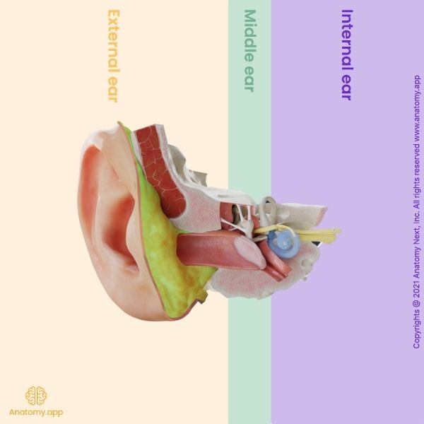
The external ear (also outer ear, auricular region of the head, Latin: auris externa) is the outer part of each ear consisting of the auricle and external acoustic meatus. At the deep end of the external acoustic meatus, separating the external ear from the middle ear, lies the tympanic membrane (eardrum).
The structures of the external ear participate in conducting and focusing sound vibrations to the eardrum and further to the middle ear that carries them to the inner ear.
The auricle of the external ear collects, concentrates, and amplifies sound waves and guides them into the external acoustic meatus. The angulation of the auricle is specific to catch sounds coming from the front more than coming from behind. The external acoustic meatus has two parts - inner and outer parts. The outer part is lined with hairy skin that contains sweat glands and sebaceous glands, which together create earwax. The hairs and earwax work as a protective barrier and disinfectant. The external acoustic meatus experiences a light bend where the outer cartilaginous part joins the bony thin inner part. This formation causes the outer part to go back while the inner part runs forwards. This works as a protective mechanism, so the foreign objects do not reach the tympanic membrane. The tympanic membrane is like a border between the ear canal and the middle ear.
Common external ear disorders
Like every organ in our body, also the external ear can experience problems and disorders. Because the external ear is the first step to collecting sound and guiding it further, external ear disorders sometimes may result in hearing impairment or possible hearing loss. Some of the most common disorders are cerumen impaction, foreign body occlusion, growths, infection, furuncle, and congenital malformations. To treat this, people may have to use softeners and ear irrigation. Too much ear wax can cause external otitis with pain, vertigo, tinnitus.
Cerumen impaction
This situation happens when earwax is accumulated in the ear canal, resulting in impaired sound flow to the tympanic membrane. Even though a small amount of wax is good for our ears, too much wax can cause hearing and balance problems.
Foreign bodies
This is one of the most common hearing problems in kids because young children like to put small toys and objects in their noses and ears. Unfortunately, this can cause trauma to the ear canal and the tympanic membrane. It is not advised to try and take out deeply put foreign objects at home by parents.
Growths
The most common growth on the external ear is exostoses. They are benign, skin-covered bony growths. There are no specific reasons why these may appear, but exostoses are more seen in people who are repeatedly exposed to cold water.
Infections
The external ear can also experience infections like external otitis and furuncle. External otitis is usually caused by bacteria known as pseudomonas. Infection can cause pain, swelling, discharge from the ear, itching, and possible hearing loss. External otitis is treated with antibiotics. Furuncle is an infection that starts when a hair follicle is infected. This infection is usually caused by staphylococcus aureus. If furuncle blocks the ear canal, the person can experience hearing loss.
Congenital malformations
Atresia of the external ear canal can cause hearing loss. In this situation, the ear canal has not developed. Many genetic syndromes affecting other parts of the body can also affect hearing and external ear development like Goldenhar syndrome (affected the development of the eyes, ears, bones of the skull, vertebrae), Treacher Collins syndrome (auricle dysplasia, atresia of the bony part of the external acoustic meatus), Crouzon syndrome (bilateral atresia of the external acoustic meatus). Sometimes surgery and hearing aids may be needed to treat these malformations.
Contact information
- For questions regarding business inquiries. Please contact:
- info@anatomy.app
Product
Platform
- Terms & Conditions
- Privacy policy
- Imprint
- .
Anatomy Next © 2024. All rights reserved.